
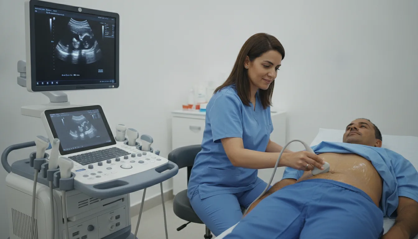
General and specialized ultrasound
Ultrasound in Tunis, Tunisia · Centre Urbain Nord
Imaging technique using ultrasound waves to explore organs and tissues in real time, without ionizing radiation, suitable for many diagnostic indications.
What is it?
Ultrasound examinations performed at the clinic
Click on an examination to find out what it is for, how to prepare and how it takes place. Ultrasound does not use X-rays. Doppler ultrasound of the blood vessels (arteries, veins, carotids…) is presented with the cardiac investigations.
Abdomen, urinary tract and prostate
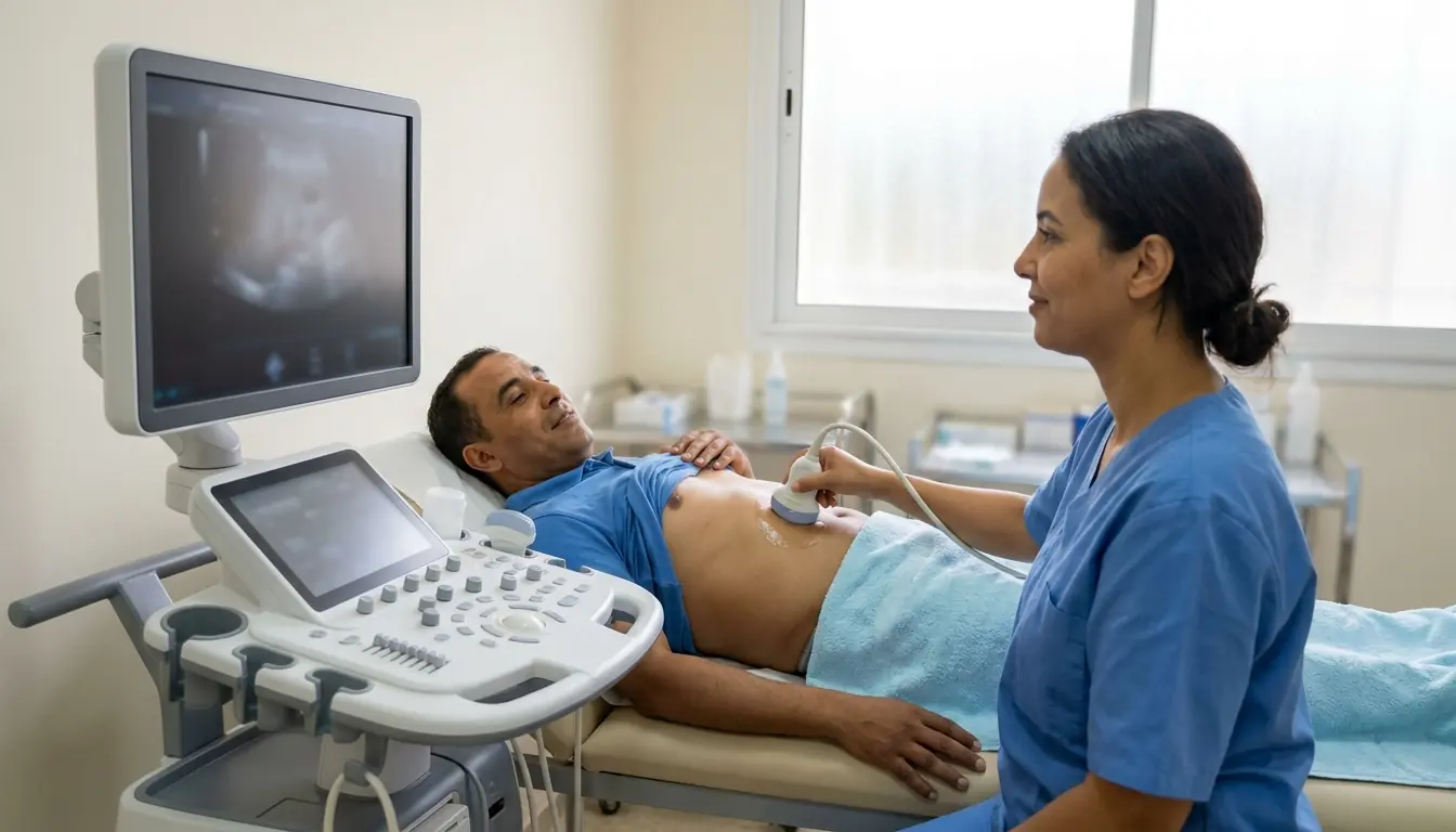
Abdominal and abdominopelvic ultrasound
Abdominal ultrasound in Tunis: liver, gallbladder, pancreas, kidneys and spleen examined with sound waves, without X-rays or injection.
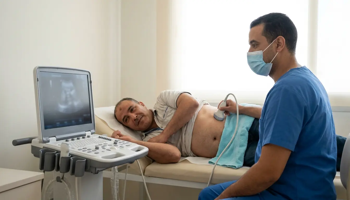
Kidney and bladder ultrasound
Kidney and bladder ultrasound in Tunis: stones, urinary infections, kidney dilatation and measurement of urine left after voiding.
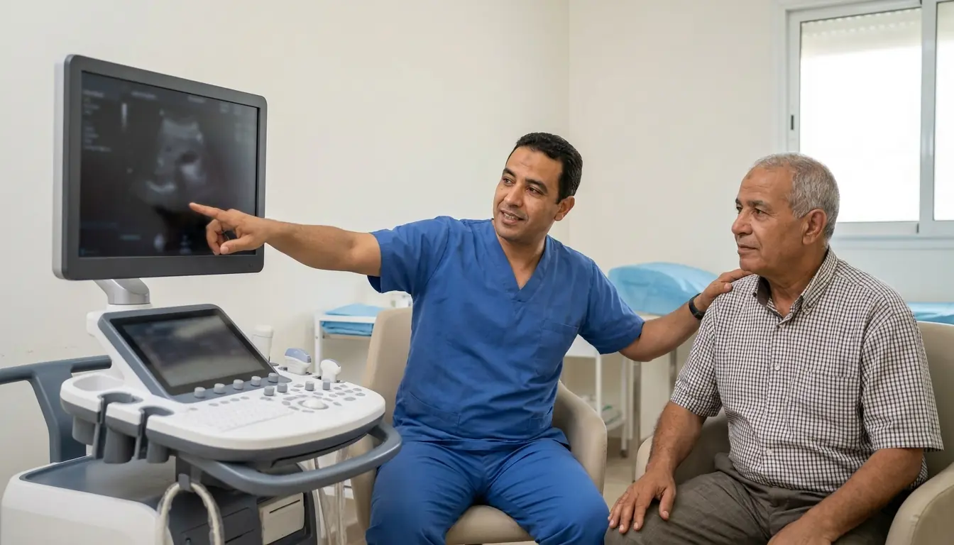
Prostate ultrasound (transabdominal or transrectal)
Prostate ultrasound in Tunis, transabdominal or transrectal: prostate volume, urinary symptoms and urine left after voiding.
Women and pregnancy
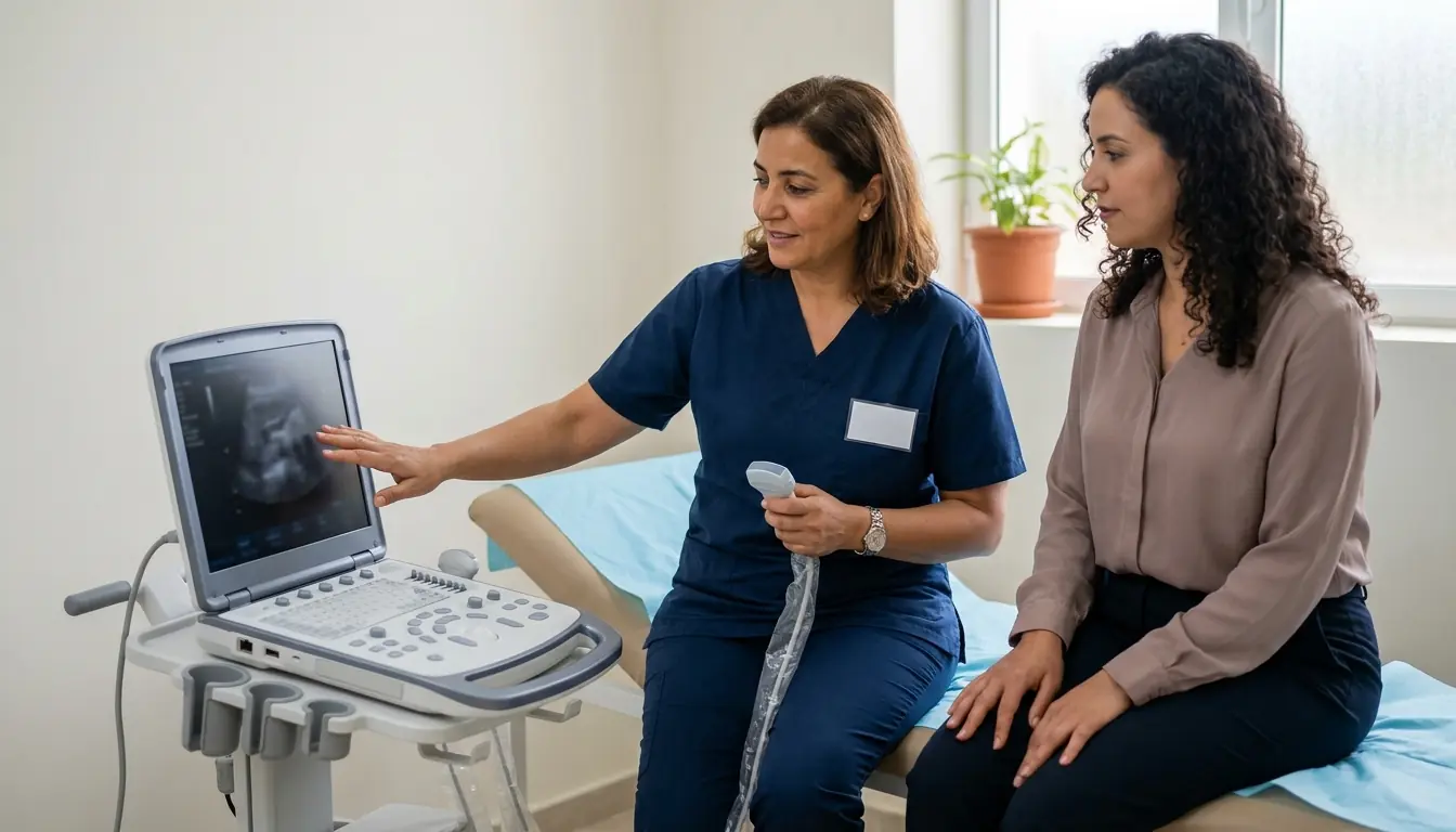
Pelvic ultrasound (transabdominal and transvaginal)
Pelvic ultrasound in Tunis: uterus and ovaries examined transabdominally or transvaginally (pain, bleeding, fibroids, cysts, fertility).
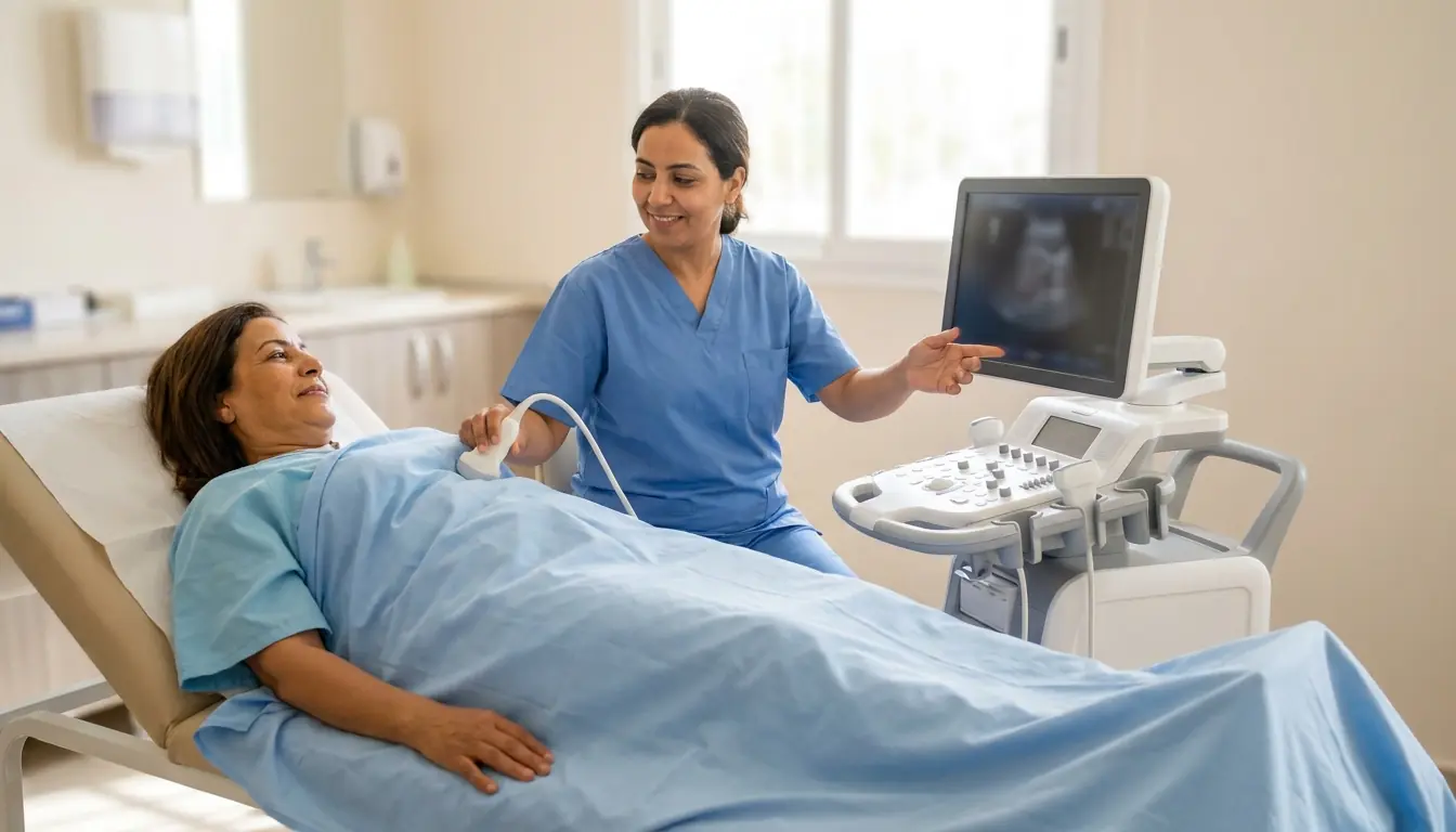
Breast ultrasound (and galactography)
Breast ultrasound in Tunis: complement to mammography, palpable lump, dense breasts, young women and follow-up after breast surgery.
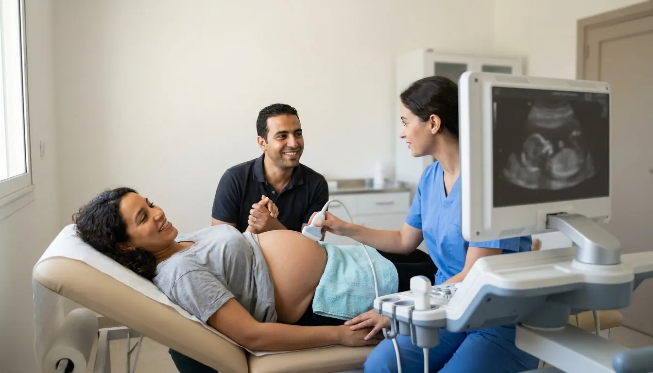
Pregnancy ultrasounds (1st, 2nd and 3rd trimester)
Pregnancy ultrasounds in Tunis: dating, nuchal translucency, baby's growth, placenta and amniotic fluid, in all three trimesters.
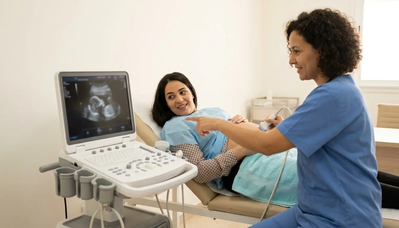
Anomaly scan (and obstetric Doppler)
Anomaly scan in Tunis: detailed study of the baby's anatomy, with obstetric Doppler if needed, for single or multiple pregnancies.
Neck, chest and soft tissues
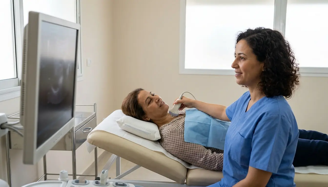
Thyroid and neck ultrasound
Thyroid ultrasound in Tunis: nodules, goitre, thyroiditis and neck lymph nodes examined with sound waves, without X-rays.
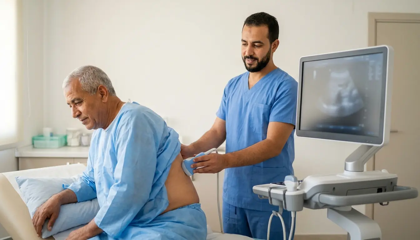
Chest ultrasound (pleura)
Chest ultrasound in Tunis: looking for pleural effusion and marking the site before drainage, with sound waves, without X-rays.
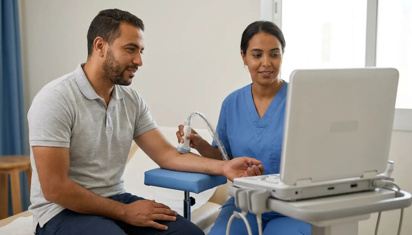
Soft tissue ultrasound (lumps, cysts, lymph nodes, hernias)
Soft tissue ultrasound in Tunis: lump under the skin, cyst, lipoma, lymph node or abdominal wall hernia examined with sound waves.
Testicular and scrotal ultrasound
Testicular ultrasound in Tunis: pain, lump, varicocele or infertility work-up, using sound waves, without X-rays.
Tendons, muscles and joints
When is it indicated?
For abdominal pain, ultrasound is often the first examination requested: it painlessly visualizes the main organs of the abdomen and looks, for example, for gallstones or an abnormality of the liver or kidneys.
If the thyroid gland appears enlarged or if a small lump is felt in the neck, ultrasound determines its size and appearance and helps decide on the appropriate course of action.
Ultrasound complements mammography, particularly in women with dense breasts. It helps distinguish a fluid-filled cyst from a solid nodule and guides any sampling procedure.
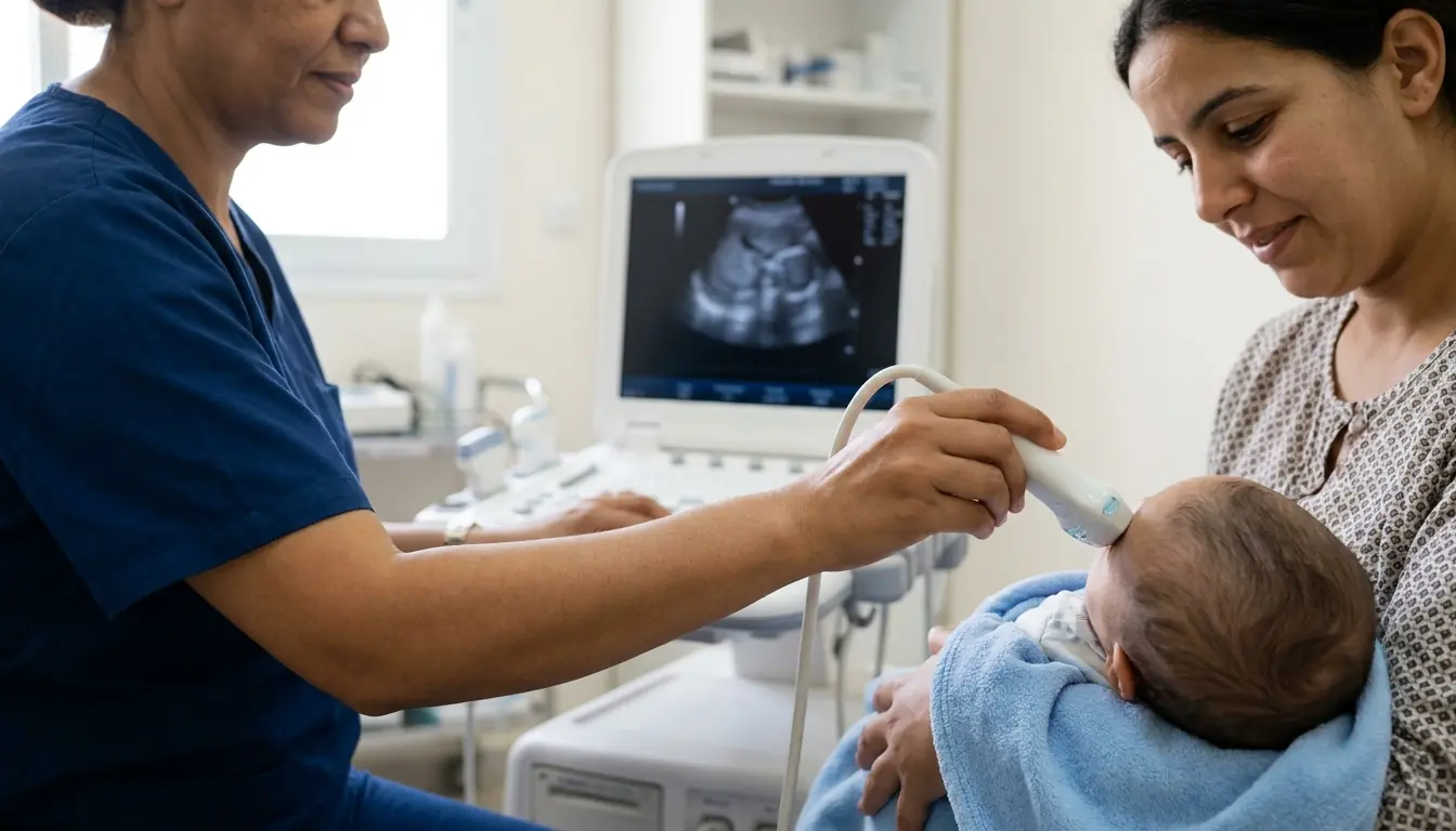
Throughout pregnancy, ultrasound monitors the baby's growth and well-being in complete safety, without any radiation, for both mother and child.
To investigate lower abdominal pain, unusually heavy periods or urinary problems, ultrasound examines the uterus, ovaries or prostate simply and painlessly.
Thanks to Doppler mode, ultrasound observes blood flowing through the vessels: it looks for a clot in a swollen leg, checks the arteries of the neck or monitors the diameter of the aorta.
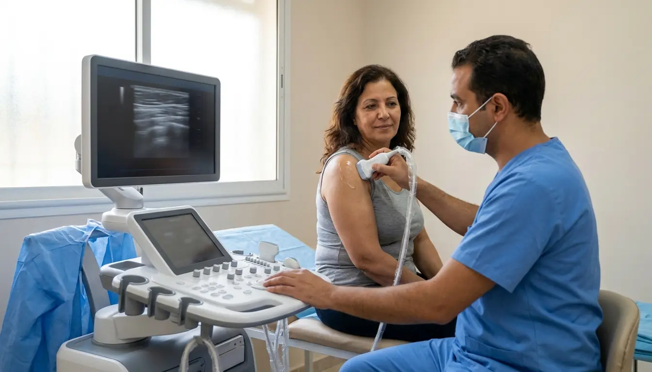
After exertion or a sports injury, ultrasound examines muscles and tendons in motion, making it possible to confirm a tear or inflammation and adapt the return to activity.
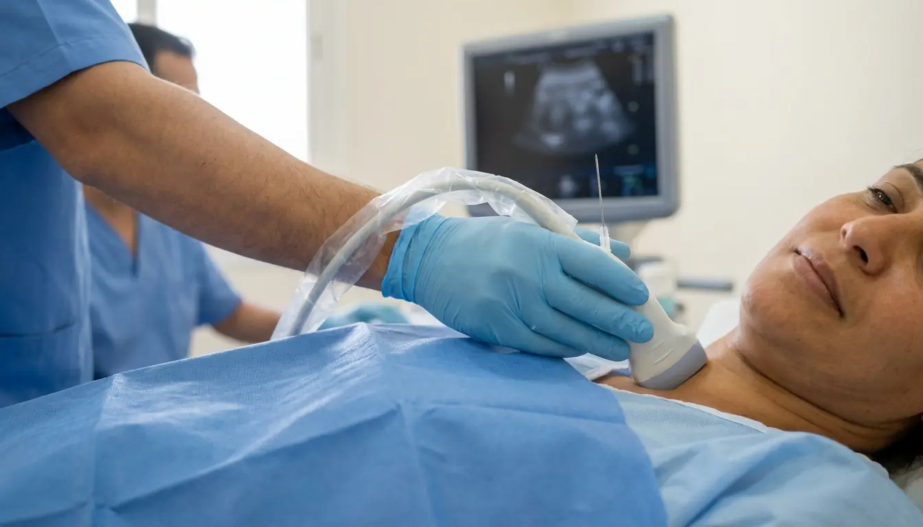
By visualizing the needle in real time, ultrasound allows the doctor to perform samplings or injections with great precision and maximum safety.
Preparation
Procedure
The patient is comfortably positioned on the examination table, generally lying on their back
Ultrasound gel is applied to the skin over the area to be explored
The radiologist applies the probe and moves it gently to visualize the different structures
The images appear in real time on the screen
The patient may be asked to change position (lying on their side, standing) to optimize the examination
Measurements and annotations are made during the examination
In Doppler mode, blood flow is analyzed with color coding
The gel is wiped off at the end of the examination
Duration
15 to 30 minutes depending on the type of ultrasound
Results
The radiologist communicates the initial results to the patient immediately after the examination. A detailed report with images is written and sent to the referring physician within 24 to 48 hours.
Risks and side effects
These risks remain rare. Your physician will inform you in detail before the examination.
Frequently asked questions
How do I book an ultrasound in Tunis?
Call the radiology centre's direct line on +216 36 402 036, or request a quote online, at Centre Urbain Nord, Tunis.
Do I need to fast before an ultrasound?
Only for abdominal ultrasound: fast for 6 hours, with a little water allowed. Other ultrasounds (pelvic, thyroid, Doppler, soft tissue) do not require fasting; the team confirms the instructions when you book.
Is ultrasound risky?
No known risk: ultrasound uses no ionising radiation. The exam is painless, takes 15 to 30 minutes and can be repeated, including during pregnancy and in children.
When are ultrasound results available?
The radiologist gives you the first results immediately after the exam. The detailed report with images is sent to your doctor within 24 to 48 hours.
Request a quote
Request your quote online or call us on the number below:
Request a Quote +216 36 402 036At a glance
Related searches
- ultrasound Tunis
- ultrasound Tunisia
- doppler ultrasound Tunis