
Abdominal and abdominopelvic ultrasound
Abdominal ultrasound in Tunis, Tunisia · Centre Urbain Nord
Abdominal ultrasound in Tunis: liver, gallbladder, pancreas, kidneys and spleen examined with sound waves, without X-rays or injection.
What is it?
When is it indicated?
It looks for a visible cause: a stone, an inflamed gallbladder, an abnormal organ or fluid.
Ultrasound is the simplest test to see gallstones and any inflammation of the gallbladder.
It examines the appearance of the liver (fatty liver, nodule) and the bile ducts.
It measures these organs and looks for a cyst, a stone or dilatation.
Without X-rays, it can be repeated to follow a lesion or an organ over time.
Preparation
Procedure
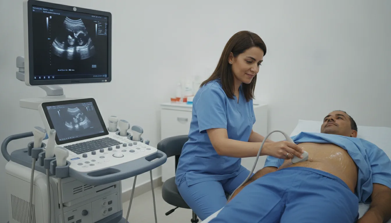
You lie on your back with your abdomen uncovered
A slightly cold gel is applied to the skin so that the ultrasound waves pass through well
The doctor moves a probe over the skin, pressing lightly; the images appear live on the screen
You may be asked to breathe in deeply, hold your breath for a few seconds or turn onto your side
The gel is wiped off; you can return to your usual activities straight away
Duration
About 15 to 30 minutes
Results
The images are analysed by the doctor performing the examination, who writes a report. The report is given to you with the relevant images and sent to your doctor, who will explain the results and the next steps. You will be told when to expect it on the day of the examination.
Precautions and contraindications
Your doctor and the radiology team will inform you in detail before the examination. Feel free to ask them your questions.
This information is general and does not replace the advice of your doctor, who chooses the examination suited to your situation.
Related searches
- Abdominal ultrasound Tunis
- Abdominal ultrasound Tunisia
- Abdominal ultrasound in Tunisia