
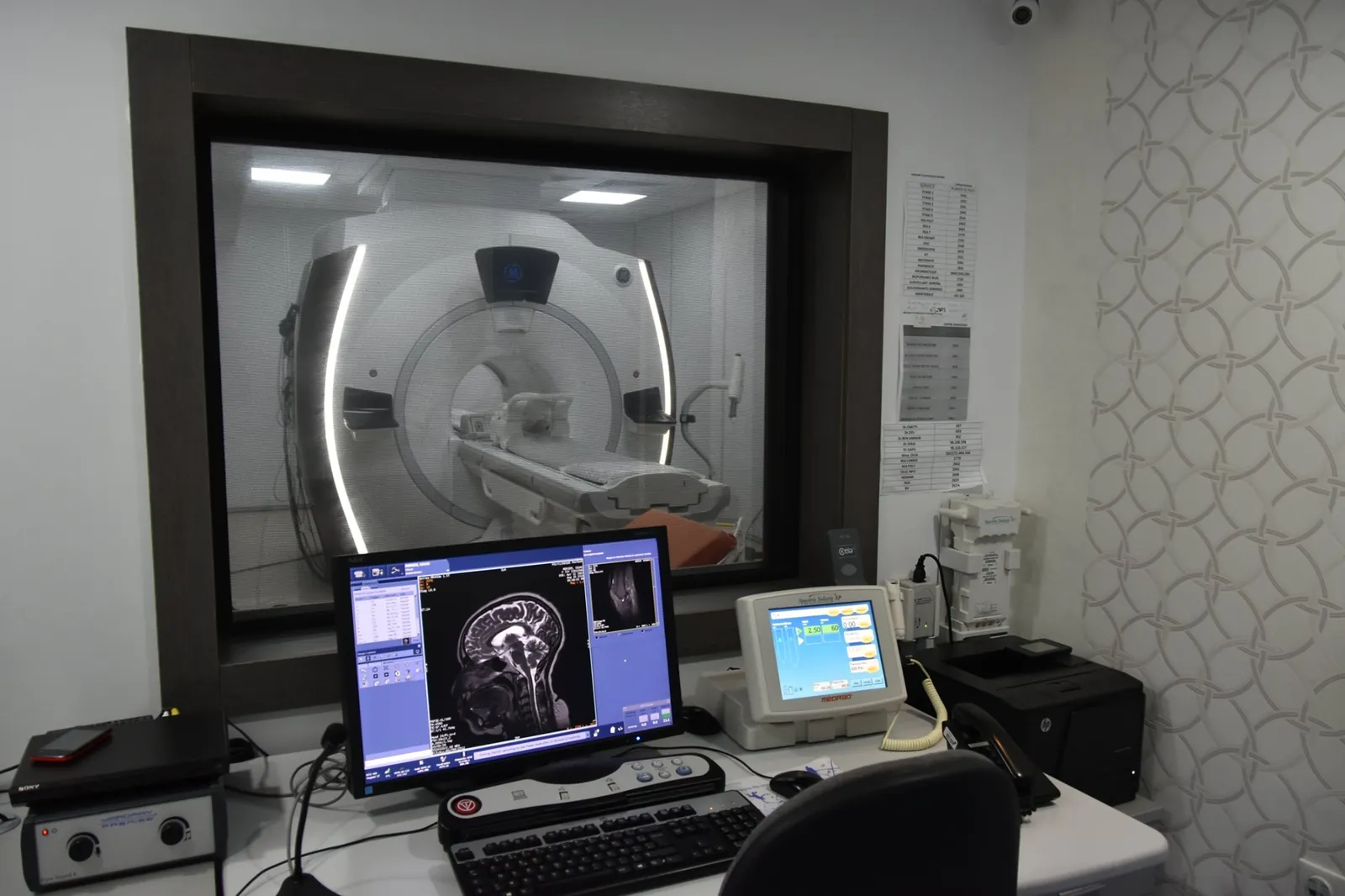
Two cross-sectional imaging exams, two different techniques
MRI (magnetic resonance imaging) and CT (computed tomography, often simply called a scanner) have one thing in common: they produce images of the body in « slices », which the radiologist can then study from every angle. That is why they are often confused. Yet they work in very different ways.
MRI uses a powerful magnetic field and radio waves. Hydrogen atoms, which are abundant in the body's tissues, react to this field and emit signals that a computer turns into images. MRI does not use any X-rays.
CT, on the other hand, uses X-rays. A tube rotates around you inside a wide ring while the table moves forward, and the computer reconstructs highly detailed images from the rays that have passed through the body. At the Clinique Pasteur radiology centre, CT exams are performed on a dual-energy scanner, a technology that analyses tissues at two X-ray energy levels.
What sets them apart in practice
For the patient, several differences are noticeable on the day of the exam:
- Radiation: MRI involves no ionising radiation; CT uses X-rays, at a dose adapted to each exam.
- Duration: a CT scan is quick, usually 10 to 20 minutes in total, with only a few seconds per pass; an MRI generally takes 20 to 45 minutes depending on the area examined.
- The machine: for an MRI you lie inside a tunnel; for a CT scan you pass through a wide, open ring.
- Noise: MRI produces loud, repetitive sounds, which is why earplugs or headphones are provided; CT is much quieter.
- Contrast agent: when an injection is needed, MRI uses gadolinium and CT uses an iodinated contrast agent.
- What they show best: MRI is particularly suited to soft tissues (brain, spinal cord, ligaments, cartilage); CT performs very well for bones, lungs and rapid examination of the whole body.
Why your doctor chooses an MRI
MRI is often preferred when tissues of similar nature need to be distinguished precisely, or when X-rays are best avoided. It is requested in particular in the following situations:
- The brain: unusual headaches, dizziness, memory problems, follow-up of certain neurological conditions.
- The spine: persistent back or neck pain, pain radiating down an arm or a leg (looking for a herniated disc, for example).
- Joints: after a sprain or a sports injury, to see the menisci, ligaments and cartilage that an X-ray does not show.
- The liver and bile ducts, as well as the pelvic organs: uterus, ovaries, prostate, rectum.
- The heart and blood vessels, using specific techniques such as magnetic resonance angiography.
Why your doctor chooses a CT scan
Because it is so fast, CT is the exam of choice when a quick assessment is needed, or when a large part of the body must be examined in one go. It is requested in particular in the following cases:
- Emergencies: after an accident, when there are signs of a stroke, or sudden abdominal pain.
- The chest: persistent cough, breathlessness, an abnormality seen on a chest X-ray.
- The abdomen: abdominal pain, an abnormality found on ultrasound, looking for an infection.
- Urinary stones: CT detects them reliably, and dual-energy technology can help determine their composition.
- Bones: complex fractures that are difficult to assess on a plain X-ray.
- Blood vessels and the heart: CT angiography, coronary CT to examine the heart's arteries without an invasive procedure.
- The assessment and follow-up of many cancers.
Preparing: what is common and what differs
In both cases, bring your prescription and any previous imaging, and report any pregnancy or possible pregnancy. Depending on the exam and whether an injection is planned, you may be asked to fast: follow the precise instructions given to you when you book.
Before an MRI, the key question is metal, because the machine is a powerful magnet. Remove jewellery, watches and piercings, and be sure to report any implant: pacemaker, prosthesis, surgical clips, stent or heart valve. Some devices, particularly older pacemakers, are not compatible with MRI. If you suffer from claustrophobia, say so when booking: the team can offer a suitable solution.
Before a CT scan with injection, report any allergy, especially to iodinated contrast agents, any kidney disease and your current medication, in particular metformin for people with diabetes. A recent blood test of kidney function may be requested. Your doctor and the radiology team will tell you, case by case, whether any treatment needs to be adjusted.
On the day of the exam and the results
Both exams are painless. You lie down, usually on your back, and the only important instruction is to stay still. During a CT scan you may sometimes be asked to hold your breath for a few seconds; a brief feeling of warmth during the injection is normal. During an MRI, an intercom lets you speak to the team at any time.
The images are then analysed by a radiologist, who writes a report sent to your doctor. Your doctor explains the results and the next steps. The time needed to receive the report depends on the type of exam; in an emergency, results are communicated to the doctor promptly.
The Clinique Pasteur radiology centre, in the Centre Urbain Nord in Tunis, performs MRI and CT scans, as well as ultrasound, mammography and conventional radiology. To book an appointment, you can call the centre directly on +216 36 402 036.
Frequently asked questions
Is an MRI more accurate than a CT scan?
Neither is superior in every situation. MRI shows soft tissues (brain, spinal cord, ligaments, cartilage) in finer detail, while CT performs very well for bones, lungs and emergencies. The right exam is the one that answers the question your doctor is asking.
Can I choose between an MRI and a CT scan myself?
The choice is made by your doctor, together with the radiologist, based on your symptoms, the area to be examined and your history (implants, allergies, kidney function, pregnancy). If you have a question or a possible contraindication, discuss it with your doctor before the exam.
Can I have an MRI with a prosthesis or a pacemaker?
It depends on the type of implant. Many recent prostheses and implants are MRI-compatible, but some devices, particularly older pacemakers, are not. Always report your implant when booking and bring its card or identification document if you have one.
Is a CT scan possible during pregnancy?
CT uses X-rays, so it is generally avoided during pregnancy. Report any pregnancy or possible pregnancy before any imaging exam, MRI included, so that your doctor can choose the exam best suited to your situation.
Is an injection always necessary?
No. Many exams are performed without an injection. When a contrast agent helps to see blood vessels or certain lesions more clearly, gadolinium is used for MRI and an iodinated agent for CT. The team will tell you in advance whether your exam includes one and how to prepare.
This article is for general information only. It does not replace the advice of your doctor, who alone can adapt instructions to your situation.
Learn more
A question? A quote?
Our team answers your questions and supports you throughout your care journey.