
Pelvic ultrasound (transabdominal and transvaginal)
Centres/Radiology/Ultrasound/Pelvic ultrasound

Pelvic ultrasound (transabdominal and transvaginal)

Pelvic ultrasound in Tunis, Tunisia · Centre Urbain Nord

Pelvic ultrasound in Tunis: uterus and ovaries examined transabdominally or transvaginally (pain, bleeding, fibroids, cysts, fertility).

What is it?

A pelvic ultrasound examines the uterus, its inner lining (endometrium) and the ovaries. It is first done through the lower abdomen (transabdominal route), with a full bladder. It is often completed by a transvaginal route: a thin probe, covered with a single-use protective sheath, is placed in the vagina to obtain more precise images, with your consent. The transvaginal route is not used in girls or women who have never had sexual intercourse: the doctor then chooses another route depending on the situation. As part of a fertility work-up, ultrasound can also be used to monitor ovulation: repeated scans during the cycle follow the growth of the follicles in the ovaries, as prescribed by the gynaecologist.

When is it indicated?

Pelvic pain

It looks for an ovarian cyst, a fibroid or another gynaecological cause.

Heavy periods or abnormal bleeding

It examines the uterus and endometrium (polyp, fibroid, thickening).

Fibroids and ovarian cysts

It measures these lesions and allows them to be monitored over time.

Fertility and ovulation monitoring

It assesses the uterus and ovaries, then follows the growth of the follicles during treatment.

Checking an IUD

It checks that the device is correctly positioned in the uterus.

Preparation

1For the transabdominal route, come with a full bladder (drink about one litre of water in the hour before, without passing urine)
2For the transvaginal route, you empty your bladder just before that part of the examination
3Tell us if you might be pregnant, the date of your last period and whether you have an IUD (coil)
4For ovulation monitoring, come on the days of the cycle given by your gynaecologist
5Bring your referral, previous imaging (ultrasound, X-rays, CT scan, MRI) and any relevant reports

Procedure

1

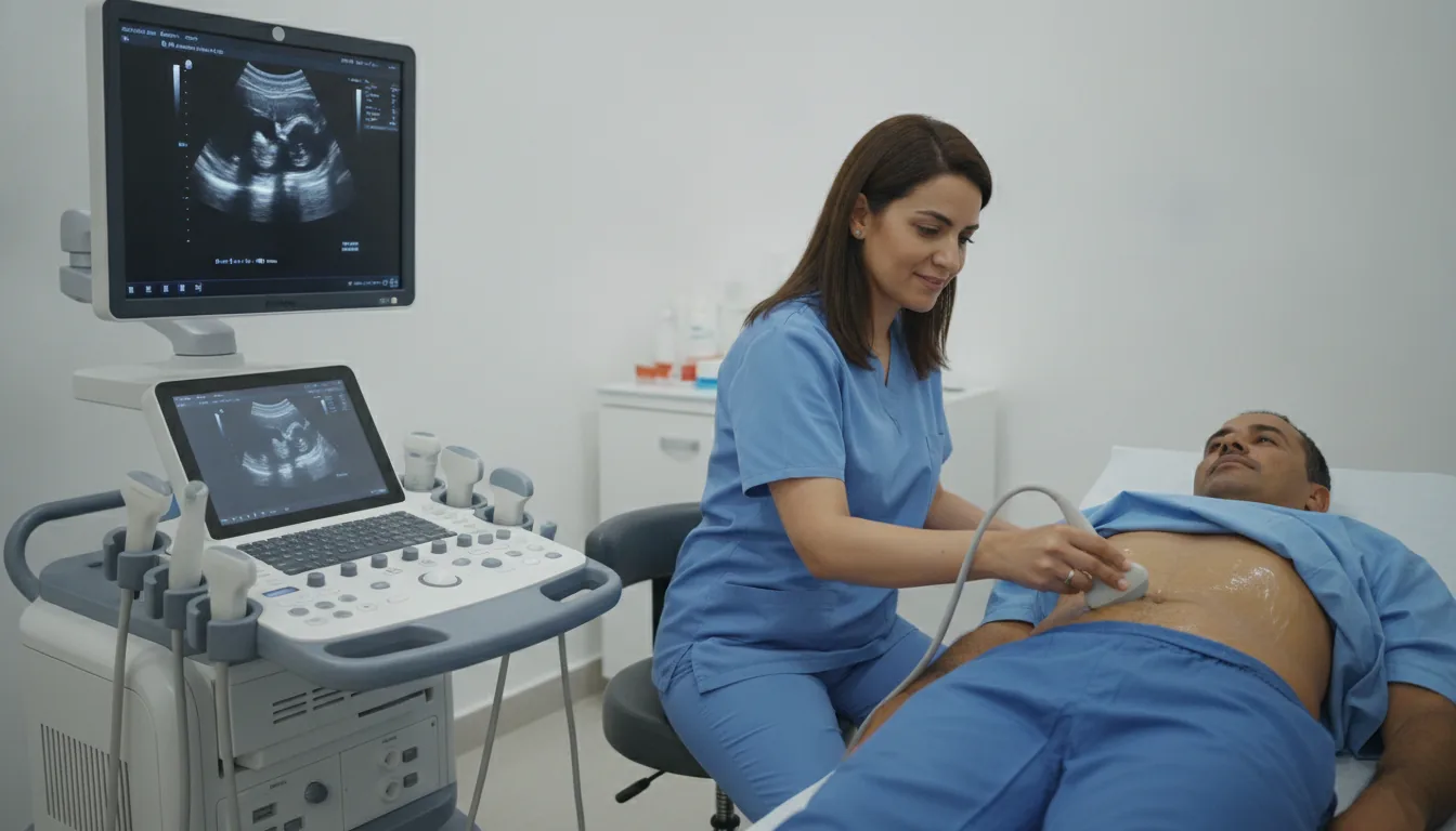
You lie on your back; the probe is first moved over the lower abdomen with a gel

2

If a transvaginal scan is useful, the doctor explains it and asks for your consent

3

The thin transvaginal probe, protected by a single-use sheath, is inserted gently; discomfort is usually mild

4

You can ask to stop at any time

5

The gel is wiped off; you can return to your usual activities straight away

Duration

About 15 to 30 minutes

Results

The images are analysed by the doctor performing the examination, who writes a report. The report is given to you with the relevant images and sent to your doctor, who will explain the results and the next steps. You will be told when to expect it on the day of the examination.

Precautions and contraindications

No X-rays: ultrasound uses sound waves, without ionising radiation
No known risk, including during pregnancy
Transvaginal route: mild discomfort possible, usually without significant pain
No contraindications: the examination can be repeated as often as needed

Your doctor and the radiology team will inform you in detail before the examination. Feel free to ask them your questions.

This information is general and does not replace the advice of your doctor, who chooses the examination suited to your situation.

Related searches

  • Pelvic ultrasound Tunis
  • Pelvic ultrasound Tunisia
  • Pelvic ultrasound in Tunisia
Request a quoteBook an appointment