
Ultrasound of tendons, muscles and joints
Musculoskeletal ultrasound in Tunis, Tunisia · Centre Urbain Nord
Musculoskeletal ultrasound in Tunis: shoulder, knee, tendinitis, muscle tears and joint effusions, with a scan in motion.
What is it?
When is it indicated?
It looks for tendinitis, calcification or a tear of the rotator cuff tendons.
It shows thickening or a tear of a tendon (elbow, wrist, knee, ankle).
After exertion, it shows the extent of the injury and helps plan the return to activity.
It finds fluid in a joint or a cyst, and can guide an aspiration.
Preparation
Procedure
You sit or lie down depending on the joint being examined
A slightly cold gel is applied to the skin so that the ultrasound waves pass through well
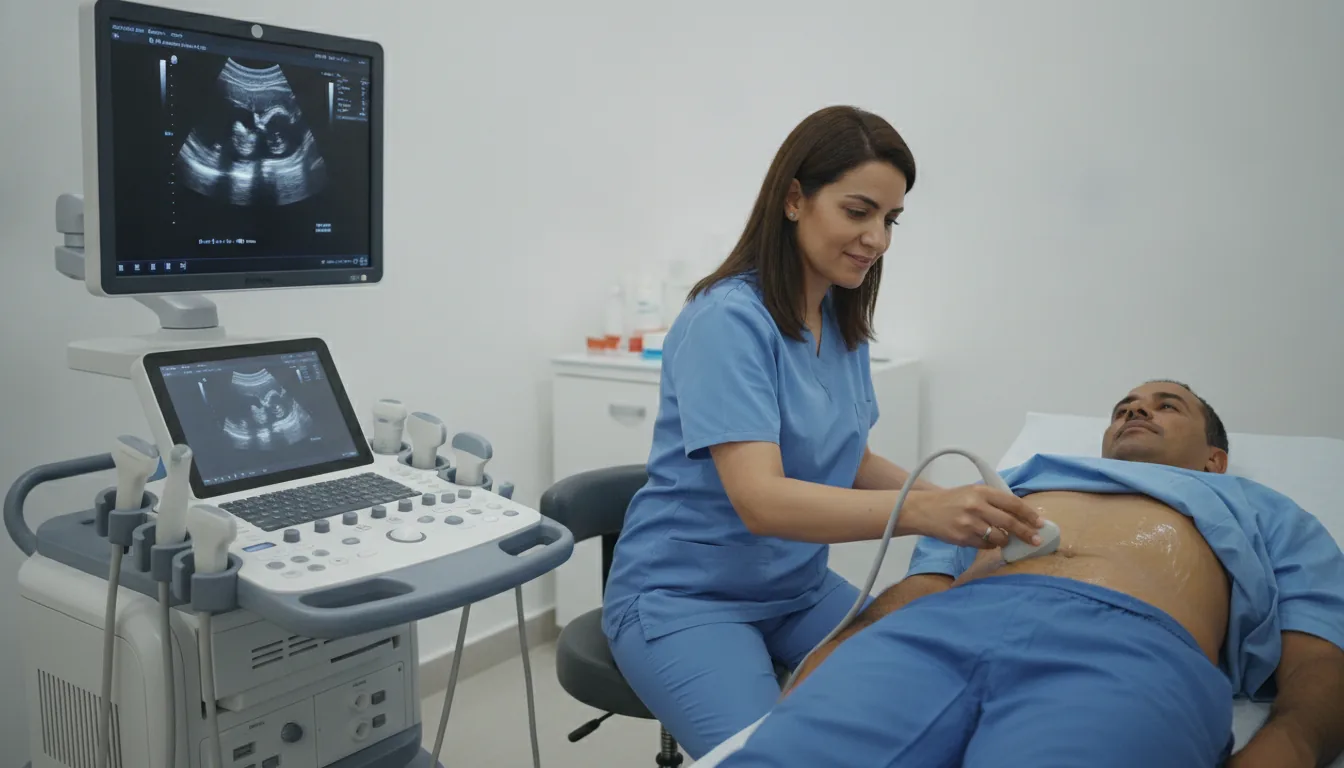
The doctor moves a probe over the skin, pressing lightly; the images appear live on the screen
You are asked to move the joint (raise your arm, bend your knee…) so that the tendons can be seen in motion
The gel is wiped off; you can return to your usual activities straight away
Duration
About 15 to 30 minutes
Results
The images are analysed by the doctor performing the examination, who writes a report. The report is given to you with the relevant images and sent to your doctor, who will explain the results and the next steps. You will be told when to expect it on the day of the examination.
Precautions and contraindications
Your doctor and the radiology team will inform you in detail before the examination. Feel free to ask them your questions.
This information is general and does not replace the advice of your doctor, who chooses the examination suited to your situation.
Related searches
- Musculoskeletal ultrasound Tunis
- Musculoskeletal ultrasound Tunisia
- Musculoskeletal ultrasound in Tunisia