
Ultrasound-guided biopsies, aspirations, injections and localisation
Centres/Radiology/Ultrasound/Ultrasound-guided procedures

Ultrasound-guided biopsies, aspirations, injections and localisation

Ultrasound-guided procedures in Tunis, Tunisia · Centre Urbain Nord

Ultrasound-guided procedures in Tunis: biopsy, aspiration, joint injection, hookwire placement or marking before surgery, under local anaesthetic.

What is it?

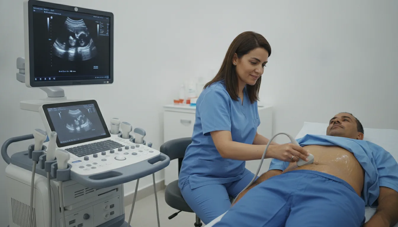
Ultrasound shows the tip of a needle live and guides it precisely to the area to be treated or sampled. These procedures are performed by the doctor, usually under local anaesthetic: - biopsy: removal of a small piece of a nodule or lump (breast, thyroid, liver, lymph node…) for laboratory analysis; - aspiration: drawing off fluid from a cyst or an effusion, to analyse it or relieve symptoms; - injection: a medicine is injected next to a tendon or into a painful joint; - hookwire placement: a very thin wire is placed in a small breast lesion to guide the surgeon during the operation; - localisation: marking a lesion or the puncture site on the skin before a procedure or surgery. The procedure is explained beforehand and your consent is obtained. The result of a biopsy is sent after laboratory analysis.

When is it indicated?

Nodule or lump to be analysed

A biopsy or fine-needle aspiration shows the nature of a lesion (breast, thyroid, liver, lymph node…).

Cyst or effusion

Aspiration drains the fluid to relieve symptoms and to analyse it.

Tendinitis or painful joint

A guided injection places the medicine exactly where it is needed.

Localisation before breast surgery

The hookwire guides the surgeon to a lesion too small to be felt.

Preparation

1Be sure to mention any anticoagulant or antiplatelet treatment (aspirin, clopidogrel…): it may need to be adjusted before the procedure, only on the advice of the prescribing doctor
2A blood test (clotting tests) may be requested before some procedures
3Tell us about any allergy (local anaesthetics, disinfectants, dressings)
4Arrange for someone to accompany you if the doctor advises it
5Bring your referral, previous imaging (ultrasound, X-rays, CT scan, MRI) and any relevant reports

Procedure

1

The doctor first locates the area with ultrasound

2

The skin is disinfected and a local anaesthetic is given

3

The needle is guided live on the screen; it is important to keep still

4

A dressing is applied; a short period of observation may follow

5

You are given instructions for the following days (taking it easy, checking the dressing)

Duration

About 20 to 45 minutes depending on the procedure, including observation

Results

The doctor explains how the procedure went and writes a report. The result of a biopsy or fluid analysis is available after laboratory examination and sent to your doctor, who will explain it to you. You will be told when to expect it on the day of the procedure.

Precautions and contraindications

No X-rays: ultrasound uses sound waves, without ionising radiation
Temporary pain at the puncture site, relieved by a simple painkiller
Minor bleeding or bruising, usually harmless
Infection: rare, thanks to disinfection and sterile equipment
Brief feeling of faintness possible during the procedure

Your doctor and the radiology team will inform you in detail before the examination. Feel free to ask them your questions.

This information is general and does not replace the advice of your doctor, who chooses the examination suited to your situation.

Related searches

  • Ultrasound-guided procedures Tunis
  • Ultrasound-guided procedures Tunisia
  • Ultrasound-guided procedures in Tunisia
Request a quoteBook an appointment