
Chest ultrasound (pleura)
Chest ultrasound in Tunis, Tunisia · Centre Urbain Nord
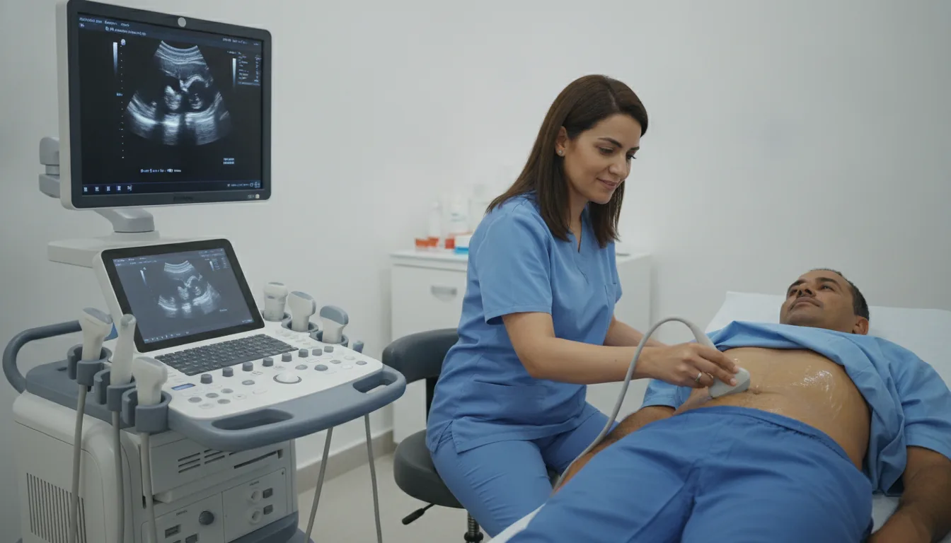
Chest ultrasound in Tunis: looking for pleural effusion and marking the site before drainage, with sound waves, without X-rays.
What is it?
When is it indicated?
It confirms fluid around the lung and estimates the amount.
It shows the safest place to sample or drain the fluid.
Without X-rays, it can be repeated to follow the effect of treatment.
Preparation
Procedure
You usually sit leaning slightly forward, with your arms crossed in front of you
A slightly cold gel is applied to the skin so that the ultrasound waves pass through well
The probe is moved over the back and sides of the chest, between the ribs
If a tap is planned afterwards, the chosen site may be marked on the skin
The gel is wiped off; you can return to your usual activities straight away
Duration
About 10 to 20 minutes
Results
The images are analysed by the doctor performing the examination, who writes a report. The report is given to you with the relevant images and sent to your doctor, who will explain the results and the next steps. You will be told when to expect it on the day of the examination.
Precautions and contraindications
Your doctor and the radiology team will inform you in detail before the examination. Feel free to ask them your questions.
This information is general and does not replace the advice of your doctor, who chooses the examination suited to your situation.
Related searches
- Chest ultrasound Tunis
- Chest ultrasound Tunisia
- Chest ultrasound in Tunisia