
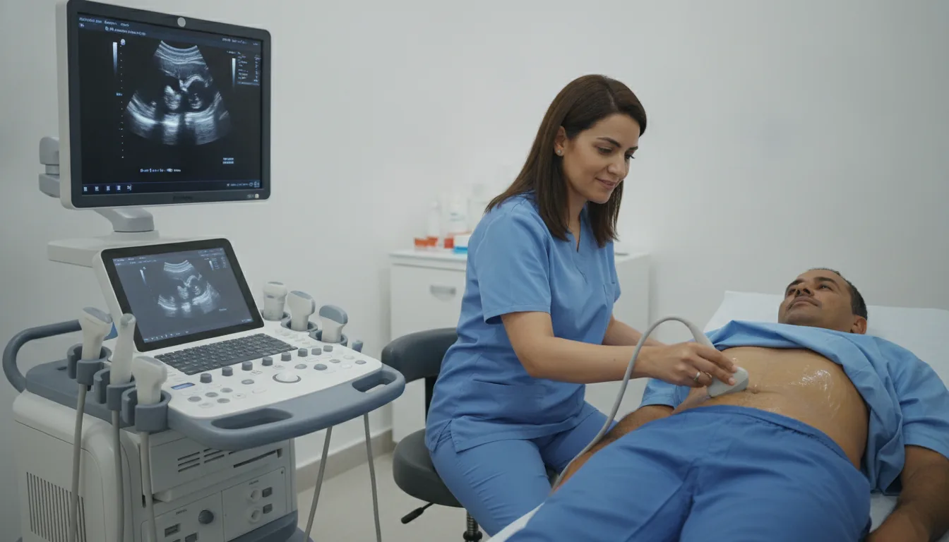
Infant ultrasounds: cranial (transfontanellar) and spinal
Infant ultrasound in Tunis, Tunisia · Centre Urbain Nord
Cranial and spinal ultrasound for infants in Tunis: the baby's brain and spinal cord examined without X-rays and without pain.
What is it?
When is it indicated?
It looks for bleeding or a brain abnormality that is common in babies born early.
It examines the brain and the fluid-filled cavities (ventricles).
Spinal ultrasound checks the position and appearance of the spinal cord.
At the paediatrician's request, it completes the baby's clinical examination.
Preparation
Procedure
The baby is settled in your arms or on the table, calmly
A lukewarm or room-temperature gel is applied to the fontanelle or to the lower back
The doctor gently places the probe without pressing; it does not hurt
You stay with your baby throughout the examination
The gel is wiped off; you can return to your usual activities straight away
Duration
About 10 to 20 minutes
Results
The images are analysed by the doctor performing the examination, who writes a report. The report is given to you with the relevant images and sent to your doctor, who will explain the results and the next steps. You will be told when to expect it on the day of the examination.
Precautions and contraindications
Your doctor and the radiology team will inform you in detail before the examination. Feel free to ask them your questions.
This information is general and does not replace the advice of your doctor, who chooses the examination suited to your situation.
Related searches
- Infant ultrasound Tunis
- Infant ultrasound Tunisia
- Infant ultrasound in Tunisia