
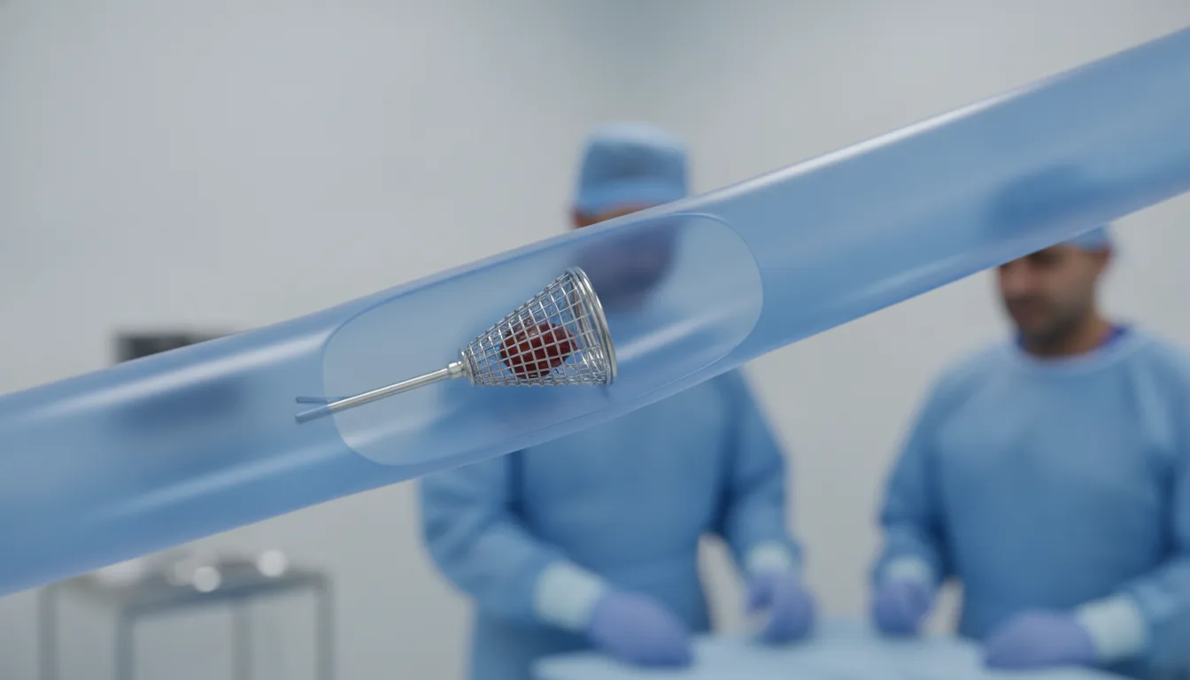
Inferior vena cava filter placement
Surgeries/Vascular/Vena cava filter

Inferior vena cava filter placement

Vena cava filter in Tunis, Tunisia · Centre Urbain Nord

Placement of a small filter in the large abdominal vein to stop a blood clot from the legs travelling to the lungs.

What is it?

The inferior vena cava is the large vein that returns blood from the legs and abdomen to the heart. When a clot forms in the leg veins (deep vein thrombosis), it can break off and travel to the lungs: this is a pulmonary embolism. A vena cava filter is a small umbrella-shaped metal device placed in this vein to catch large clots. Placement is done in the catheterisation laboratory under local anaesthesia, through a puncture of a vein in the neck or groin. The filter is opened at the right level, just below the kidney veins, under X-ray guidance. The procedure is short and requires no incision. The standard treatment for deep vein thrombosis is anticoagulation. A filter is reserved for patients who cannot receive it (bleeding, urgent surgery) or in whom it has failed. Most filters used today are retrievable (optional) and should be removed as soon as anticoagulation is possible again, through a similar procedure. At Clinique Pasteur Tunis, filter placement and removal are performed in a latest-generation catheterisation laboratory, in coordination with the doctors treating the thrombosis.
Inferior vena cava filter placement

When is it indicated?

Deep vein thrombosis or pulmonary embolism when anticoagulants cannot be used

If bleeding or surgery prevents blood thinning, the filter protects the lungs until treatment can be restarted.

Recurrent embolism despite well-managed anticoagulation

In this uncommon situation, the filter adds mechanical protection to the treatment.

Anticoagulation that has to be stopped

A bleeding complication during treatment may force it to stop; the filter then takes over temporarily.

Selected very high-risk situations, case by case

Some patients with multiple injuries or facing major surgery may benefit, after discussion between specialists.

Preparation

1Doppler ultrasound or CT confirming the thrombosis or embolism
2Blood tests including clotting and kidney function
3Report any iodine allergy
4Fast for a few hours before the procedure

Procedure

1

Positioning in the catheterisation laboratory and local anaesthesia

2

Puncture of a vein in the neck or groin

3

Imaging of the vena cava to locate the kidney veins

4

Advancement of the folded filter inside a catheter

5

Deployment of the filter at the right level and X-ray check

6

Removal of the catheter and a pressure dressing

Duration

About 30 minutes

Aftercare & Recovery

You are monitored for a few hours; the length of the hospital stay mainly depends on the condition that led to the filter. Anticoagulation is restarted as soon as possible. Removal of a temporary filter is scheduled in the following weeks or months, through a similar procedure.

Risks and side effects

Bruising at the puncture site
Clotting of the vena cava or the filter, which is why anticoagulation is resumed
Movement or tilting of the filter, rare
Removal may be more difficult if the filter is left in too long

These risks remain rare. Your surgeon will inform you in detail before the procedure.

Request a Quote

Contact us to schedule a consultation or receive a personalized quote.

Request a Quote +216 36 402 000

At a glance

Duration: About 30 minutes
Preparation: Required
Dedicated specialist team

Related searches

  • Vena cava filter Tunis
  • Vena cava filter Tunisia
  • Vena cava filter in Tunisia
Request a quote