
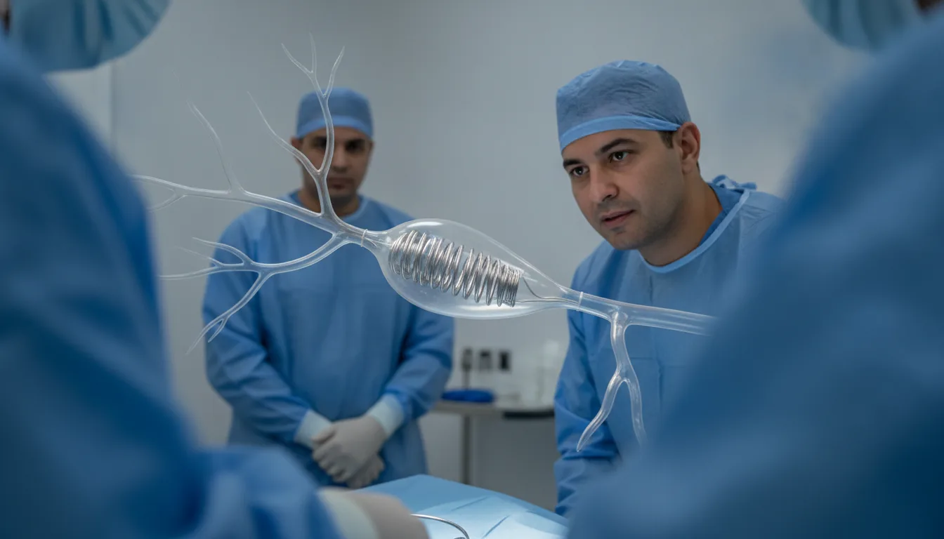
Brain aneurysm coiling
Surgeries/Vascular/Brain aneurysm (coils)

Brain aneurysm coiling

Brain aneurysm (coils) in Tunis, Tunisia · Centre Urbain Nord

Treatment of an aneurysm of a brain artery by filling it with fine platinum coils, via a catheter and without opening the skull.

What is it?

A brain aneurysm is a small pouch that forms on the wall of an artery in the brain. If it ruptures, it causes serious bleeding (subarachnoid haemorrhage). Embolisation fills the pouch from the inside with very fine platinum spirals, called coils, so that blood can no longer enter and the aneurysm is excluded from the circulation. The procedure is performed under general anaesthesia in the catheterisation laboratory. A catheter is introduced through the groin or wrist and advanced to the brain arteries, then a microcatheter is placed inside the aneurysm. Depending on its shape, the doctor may use a balloon or a stent to hold the coils in place, or other suitable devices. This technique is used as an emergency after a rupture, and as a planned procedure for some aneurysms found by chance whose risk of rupture justifies treatment. The choice between embolisation and surgery (clipping) depends on the shape and position of the aneurysm; it is discussed between interventional neuroradiologists and neurosurgeons. At Clinique Pasteur Tunis, the procedure is performed in a latest-generation catheterisation laboratory, with neurosurgery and intensive care on site.
Brain aneurysm coiling

When is it indicated?

Ruptured aneurysm (subarachnoid haemorrhage)

After a rupture, the aneurysm must be closed quickly to prevent further bleeding, which would be very serious.

Unruptured aneurysm at risk of rupture

Depending on its size, shape and location, an aneurysm found by chance may warrant preventive treatment.

Growing or symptomatic aneurysm

An increase in size or pressure on a nerve (drooping eyelid, double vision) leads to treatment.

Aneurysm more accessible from inside the artery

Some deep locations are easier and safer to treat by the endovascular route than by surgery.

Preparation

1CT, MRI or cerebral angiography to assess the aneurysm
2Shared decision between the interventional neuroradiologist and the neurosurgeon
3Anaesthetic consultation and blood tests
4Antiplatelet treatment started beforehand if a stent is planned
5Fast for 6 hours before the procedure (except in an emergency)

Procedure

1

General anaesthesia in the catheterisation laboratory

2

Puncture of the artery in the groin or at the wrist

3

Advancement of the catheter to the brain arteries

4

Placement of a microcatheter inside the aneurysm

5

Successive placement of coils, with a balloon or stent if needed

6

Check that the aneurysm is closed and waking up under monitoring

Duration

About 1.5 to 3 hours

Aftercare & Recovery

For an unruptured aneurysm, the hospital stay is usually 2 to 3 days, with a return to activities within one to two weeks. After a rupture, the length of stay depends on the haemorrhage and begins in intensive care. Follow-up imaging (MRI or angiography) is scheduled in the following months, as the aneurysm can sometimes partly reopen.

Risks and side effects

Clot forming in a brain artery, prevented by anticoagulant treatment during the procedure
Rupture of the aneurysm during the procedure, rare and managed immediately
Partial reopening of the aneurysm, which may require further treatment
Headaches in the first days, and bruising at the puncture site

These risks remain rare. Your surgeon will inform you in detail before the procedure.

Request a Quote

Contact us to schedule a consultation or receive a personalized quote.

Request a Quote +216 36 402 000

At a glance

Duration: About 1.5 to 3 hours
Preparation: Required
Dedicated specialist team

Related searches

  • Brain aneurysm (coils) Tunis
  • Brain aneurysm (coils) Tunisia
  • Brain aneurysm (coils) in Tunisia
Request a quote