
Lower limb arteriography
Surgeries/Vascular/Leg arteriography

Lower limb arteriography

Leg arteriography in Tunis, Tunisia · Centre Urbain Nord

An X-ray examination of the leg arteries, performed through a simple puncture, to pinpoint a narrowing or blockage.

What is it?

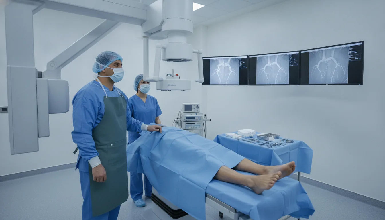
Lower limb arteriography is an examination that shows the inside of the leg arteries. An iodinated contrast dye is injected into the artery through a thin catheter, and X-ray images show in real time how the blood flows, where the arteries are narrowed (stenoses) and where they are blocked (occlusions). The examination takes place in the catheterisation laboratory under local anaesthesia. The doctor punctures an artery, usually in the groin and sometimes at the wrist, then advances the catheter under X-ray guidance. There is no incision: only a small puncture mark remains at the end. Today, Doppler ultrasound and CT scanning are often enough to make the diagnosis. Arteriography is mainly offered when treatment is being considered: it provides a very precise map of the arteries and, if needed and planned in advance, allows the artery to be widened during the same session. At Clinique Pasteur Tunis, arteriography is performed in a latest-generation catheterisation laboratory by a team experienced in endovascular procedures, with appropriate monitoring afterwards.
Lower limb arteriography

When is it indicated?

Leg pain when walking (intermittent claudication)

Cramps in the calf or thigh that force you to stop walking may be caused by narrowed arteries. Arteriography locates them precisely.

Pain at rest or a foot wound that will not heal

These signs point to a serious lack of blood in the foot. An exact map of the arteries is then needed to choose the best treatment.

Planning an angioplasty or a bypass

Before reopening or bypassing an artery, the surgeon needs to know the number, length and position of the lesions.

Inconclusive Doppler or CT results

When non-invasive tests are not enough to decide, arteriography gives a sharper view of the small arteries of the leg.

Preparation

1Prior consultation and blood tests, particularly kidney function
2Report any allergy, especially to iodine or contrast dye
3Adjust certain medicines as instructed by your doctor (anticoagulants, metformin)
4Fast for a few hours before the examination
5Drink plenty of fluids in the preceding days, unless advised otherwise

Procedure

1

Positioning on the table in the catheterisation laboratory

2

Skin disinfection and local anaesthesia in the groin or at the wrist

3

Puncture of the artery and placement of a small sheath

4

Advancement of the catheter and injection of contrast dye, with a brief feeling of warmth

5

Recording of images of the arteries, from the abdomen down to the feet

6

Removal of the equipment and compression or closure of the puncture site

Duration

About 30 to 45 minutes

Aftercare & Recovery

You stay lying down for a few hours to protect the puncture site. The examination is usually done as a day case or with one night in hospital. Strenuous effort and heavy lifting should be avoided for a few days. The results are discussed with your doctor to decide on treatment.

Risks and side effects

Bruising at the puncture site, usually minor
Allergic reaction to the contrast dye, prevented by careful questioning beforehand
Temporary effect on kidney function, monitored with blood tests
Rare arterial complications (dissection, embolism), treated immediately

These risks remain rare. Your surgeon will inform you in detail before the procedure.

Request a Quote

Contact us to schedule a consultation or receive a personalized quote.

Request a Quote +216 36 402 000

At a glance

Duration: About 30 to 45 minutes
Preparation: Required
Dedicated specialist team

Related searches

  • Leg arteriography Tunis
  • Leg arteriography Tunisia
  • Leg arteriography in Tunisia
Request a quote