
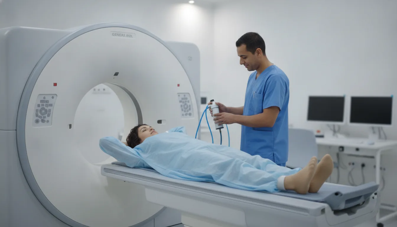
CT myelography (and myelography)
Centres/Radiology/CT scan/CT myelography

CT myelography (and myelography)

CT myelography in Tunis, Tunisia · Centre Urbain Nord

CT myelography in Tunis: CT scan after contrast is injected around the spinal cord by lumbar puncture, when MRI is not possible or not enough.

What is it?

CT myelography examines the spinal canal, the spinal cord and the nerve roots. The radiologist first injects an iodinated contrast agent into the fluid around the spinal cord, through a puncture in the lower back (lumbar puncture); X-rays are taken (myelography), followed by a CT scan. It is offered when MRI is not possible (non-compatible pacemaker, certain implants) or when it is not enough to answer the question, for example to clarify compression of a nerve root before surgery. Depending on the request, the examination covers the cervical, thoracic or lumbar region, several regions or the whole spine.

When is it indicated?

MRI not possible

With a non-compatible pacemaker or certain implants, it replaces MRI to study the spinal cord and nerves.

Nerve root compression

It clarifies a herniated disc or narrowing pressing on a nerve, before surgery.

Spinal stenosis

It shows how narrow the canal is along the whole length studied.

Assessment after spinal surgery

When metal hardware interferes with MRI, it allows the nerves to be checked.

Preparation

1Tell us if you are pregnant or think you might be: a CT scan uses X-rays
2Tell us about any blood-thinning treatment (anticoagulant, antiplatelet): whether to stop it is decided by the doctor, never on your own
3If an injection is planned, tell us about any allergy (especially to an iodinated contrast agent), kidney disease, diabetes or myeloma; a recent blood test (creatinine) may be requested
4Tell us about epilepsy and the medicines you take: some may need adjusting, according to medical advice
5Do not eat for a few hours as instructed, and arrange for someone to take you home
6Bring your referral, previous imaging (CT scan, MRI, X-rays, ultrasound) and any relevant reports

Procedure

1

You sit leaning forward or lie on your side; the skin of the lower back is disinfected and a local anaesthetic is given

2

A fine needle is inserted between two vertebrae; the contrast agent is injected into the fluid around the spinal cord

3

X-rays are taken while tilting the table, then you move to the CT scanner

4

After the examination, you rest lying down for a few hours under observation, as instructed

5

Drinking plenty and avoiding exertion for one to two days is advised

Duration

About 1 hour for the examination, followed by a few hours of rest on site

Results

The images are analysed by a radiologist, who writes a report. The report is given to you with the images and sent to your doctor, who will explain the results and the next steps. You will be told when to expect it on the day of the examination.

Precautions and contraindications

A CT scan uses X-rays: the dose is set as low as possible while still giving useful images, and the examination is only done when it is justified
Pregnancy: a relative contraindication; a CT scan is only performed if it is essential, after medical advice
Headache after the puncture, especially when standing: it usually eases with lying down and drinking; tell the team if it persists
Temporary lower back pain at the puncture site
Allergic reaction to the iodinated contrast agent (if injected): rare and usually mild (redness, itching); serious reactions are exceptional and the team is trained to manage them
Serious complications (infection, bleeding around the spinal cord, seizures): exceptional

Your doctor and the radiology team will inform you in detail before the examination. Feel free to ask them your questions.

This information is general and does not replace the advice of your doctor, who chooses the examination suited to your situation.

Related searches

  • CT myelography Tunis
  • CT myelography Tunisia
  • CT myelography in Tunisia
Request a quoteBook an appointment