
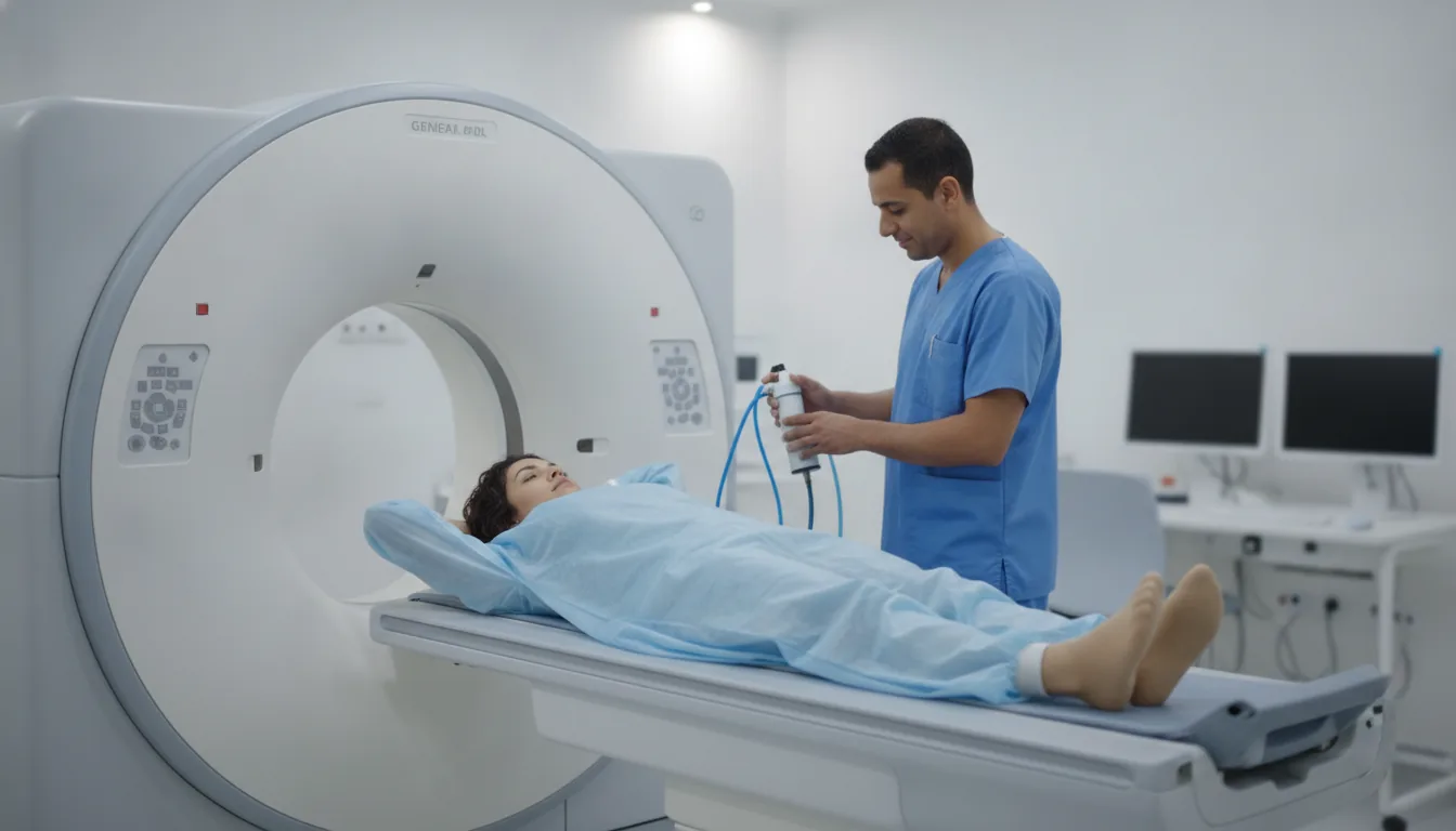
Spine CT scan (cervical, thoracic, lumbar)
Spine CT scan in Tunis, Tunisia · Centre Urbain Nord
CT scan of the cervical, thoracic or lumbar spine in Tunis: vertebrae and discs, for disc herniation, fracture, arthritis or spinal stenosis.
What is it?
When is it indicated?
When MRI is not possible, it shows a herniated disc pressing on a nerve root.
It looks for and describes a vertebral fracture after a fall or accident.
It shows bone spurs and narrowing of the canal through which the nerves pass.
It helps plan the operation or checks the position of hardware and bone healing.
Preparation
Procedure
You remove clothing and metal objects from the area to be examined; a gown may be provided
You lie on your back; a cushion under your knees eases the lower back
You lie on a table that slides slowly through a wide, open ring: it is not a closed tunnel
You need to keep still during the pass, which lasts a few seconds
The radiographer can see and hear you at all times from the next room
You can resume your activities straight away
Duration
About 10 to 15 minutes in total
Results
The images are analysed by a radiologist, who writes a report. The report is given to you with the images and sent to your doctor, who will explain the results and the next steps. You will be told when to expect it on the day of the examination.
Precautions and contraindications
Your doctor and the radiology team will inform you in detail before the examination. Feel free to ask them your questions.
This information is general and does not replace the advice of your doctor, who chooses the examination suited to your situation.
Related searches
- Spine CT scan Tunis
- Spine CT scan Tunisia
- Spine CT scan in Tunisia