
CT scan of bones and joints (pelvis, knee, ankle, wrist, extremities)
Bone and joint CT in Tunis, Tunisia · Centre Urbain Nord
CT scan of bones and joints in Tunis (pelvis, knee, ankle, wrist, hand, foot): complex fracture, assessment before surgery, bone healing.
What is it?
When is it indicated?
It shows all the fragments of a fracture, particularly in a joint, to plan surgery.
It checks that the bone has knitted properly after a fracture or surgery.
It helps plan a joint replacement or the correction of a deformity.
It clarifies a bone abnormality seen on an X-ray.
Preparation
Procedure
You lie on the table with the limb being examined positioned and supported so it does not move (sometimes with your arm above your head for the wrist)
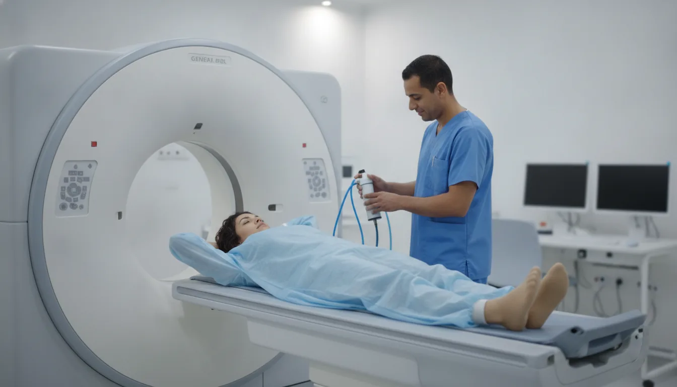
You lie on a table that slides slowly through a wide, open ring: it is not a closed tunnel
You need to keep still during the pass, which lasts a few seconds
The radiographer can see and hear you at all times from the next room
You can resume your activities straight away
Duration
About 10 to 15 minutes in total
Results
The images are analysed by a radiologist, who writes a report. The report is given to you with the images and sent to your doctor, who will explain the results and the next steps. You will be told when to expect it on the day of the examination.
Precautions and contraindications
Your doctor and the radiology team will inform you in detail before the examination. Feel free to ask them your questions.
This information is general and does not replace the advice of your doctor, who chooses the examination suited to your situation.
Other examinations — CT scan
All examinationsRelated searches
- Bone and joint CT Tunis
- Bone and joint CT Tunisia
- Bone and joint CT in Tunisia