
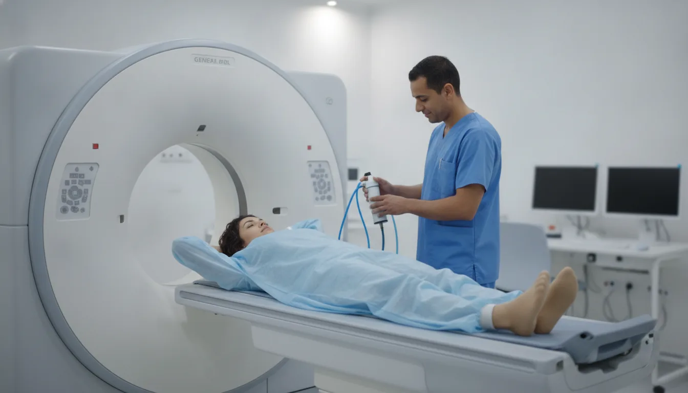
CT scan of the sinuses, facial bones and orbits
Centres/Radiology/CT scan/Sinus and face CT

CT scan of the sinuses, facial bones and orbits

Sinus and face CT in Tunis, Tunisia · Centre Urbain Nord

CT scan of the sinuses, face and orbits in Tunis: chronic sinusitis, assessment before sinus surgery, facial injury, low-dose examination.

What is it?

A CT scan of the sinuses and facial bones shows in detail the bones of the face, the sinus cavities, the nasal passages and the orbits (the bony cavities that hold the eyes). It is the test that best shows the thin bony walls of this area. It is most often requested by an ENT doctor for sinusitis that lasts or keeps coming back, a permanently blocked nose, polyps, or before sinus surgery to guide the surgeon. It is also useful after a facial injury (fracture of the nose, cheekbone, jaw or orbit). It is usually done without an injection and with a low radiation dose. Depending on the request, it can be combined with a brain CT scan or focused on the orbits.

When is it indicated?

Chronic or recurrent sinusitis

It shows which sinuses are affected and whether something is blocking their drainage.

Assessment before sinus surgery

It acts as a map for the surgeon to operate safely close to the eyes and brain.

Polyps, permanently blocked nose

It shows the extent of polyps or a deviated nasal septum.

Injury to the face or orbit

It looks for a fracture of the facial bones and guides treatment.

Preparation

1Tell us if you are pregnant or think you might be: a CT scan uses X-rays
2Remove glasses, earrings, facial piercings and removable dentures
3If the scan is for chronic sinusitis, your doctor may ask for it to be done away from a flare-up or after treatment
4If an injection is planned, tell us about any allergy (especially to an iodinated contrast agent), kidney disease, diabetes or myeloma; a recent blood test (creatinine) may be requested
5Bring your referral, previous imaging (CT scan, MRI, X-rays, ultrasound) and any relevant reports

Procedure

1

You lie on your back, with your head held in a support

2

You lie on a table that slides slowly through a wide, open ring: it is not a closed tunnel

3

You need to keep your head still and avoid swallowing during the pass, which lasts a few seconds

4

An injection is only used in certain cases (a mass, a complication of infection)

5

The radiographer can see and hear you at all times from the next room

6

You can then return to your usual activities; after an injection, drink plenty of fluids during the day (unless your doctor advises otherwise) to help clear the contrast agent

Duration

About 5 to 15 minutes in total

Results

The images are analysed by a radiologist, who writes a report. The report is given to you with the images and sent to your doctor, who will explain the results and the next steps. You will be told when to expect it on the day of the examination.

Precautions and contraindications

A CT scan uses X-rays: the dose is set as low as possible while still giving useful images, and the examination is only done when it is justified
Pregnancy: a relative contraindication; a CT scan is only performed if it is essential, after medical advice
Allergic reaction to the iodinated contrast agent (if injected): rare and usually mild (redness, itching); serious reactions are exceptional and the team is trained to manage them
In case of kidney failure, the iodinated injection can strain the kidneys: the doctor avoids or adjusts it

Your doctor and the radiology team will inform you in detail before the examination. Feel free to ask them your questions.

This information is general and does not replace the advice of your doctor, who chooses the examination suited to your situation.

Related searches

  • Sinus and face CT Tunis
  • Sinus and face CT Tunisia
  • Sinus and face CT in Tunisia
Request a quoteBook an appointment