
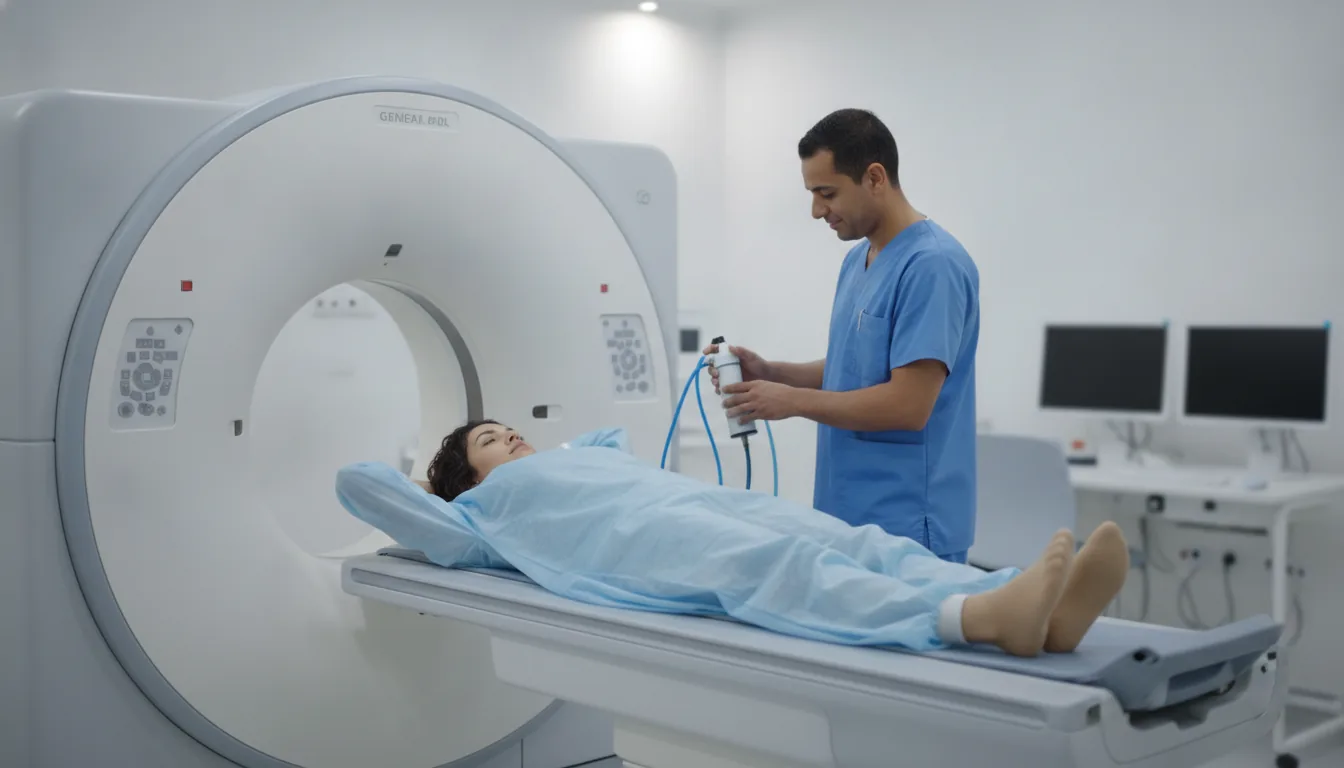
CT arthrography and arthrography
CT arthrography in Tunis, Tunisia · Centre Urbain Nord
CT arthrography and arthrography in Tunis: contrast injected into the joint, then images, for the shoulder, knee, wrist or hip.
What is it?
When is it indicated?
It shows injuries of the labrum and ligaments before possible stabilisation surgery.
It clarifies a tear of the shoulder tendons and the condition of the muscles.
It looks for a meniscal tear or cartilage injury, particularly when MRI is not possible or after surgery.
It examines the small ligaments of the wrist after a fall or with persistent pain.
It looks for a cartilage injury or a loose fragment in the joint.
Preparation
Procedure
You are positioned on the X-ray table; the skin is carefully disinfected (aseptic technique)
A local anaesthetic is given, then the radiologist inserts a fine needle into the joint under image guidance
The contrast agent is injected: a feeling of pressure or tightness in the joint is normal
You then move to the CT scanner, lying down with the limb still, for a few minutes
A dressing is applied; resting the joint for the rest of the day is advised
Duration
About 30 to 45 minutes in total (injection then CT scan)
Results
The images are analysed by a radiologist, who writes a report. The report is given to you with the images and sent to your doctor, who will explain the results and the next steps. You will be told when to expect it on the day of the examination.
Precautions and contraindications
Your doctor and the radiology team will inform you in detail before the examination. Feel free to ask them your questions.
This information is general and does not replace the advice of your doctor, who chooses the examination suited to your situation.
Related searches
- CT arthrography Tunis
- CT arthrography Tunisia
- CT arthrography in Tunisia