
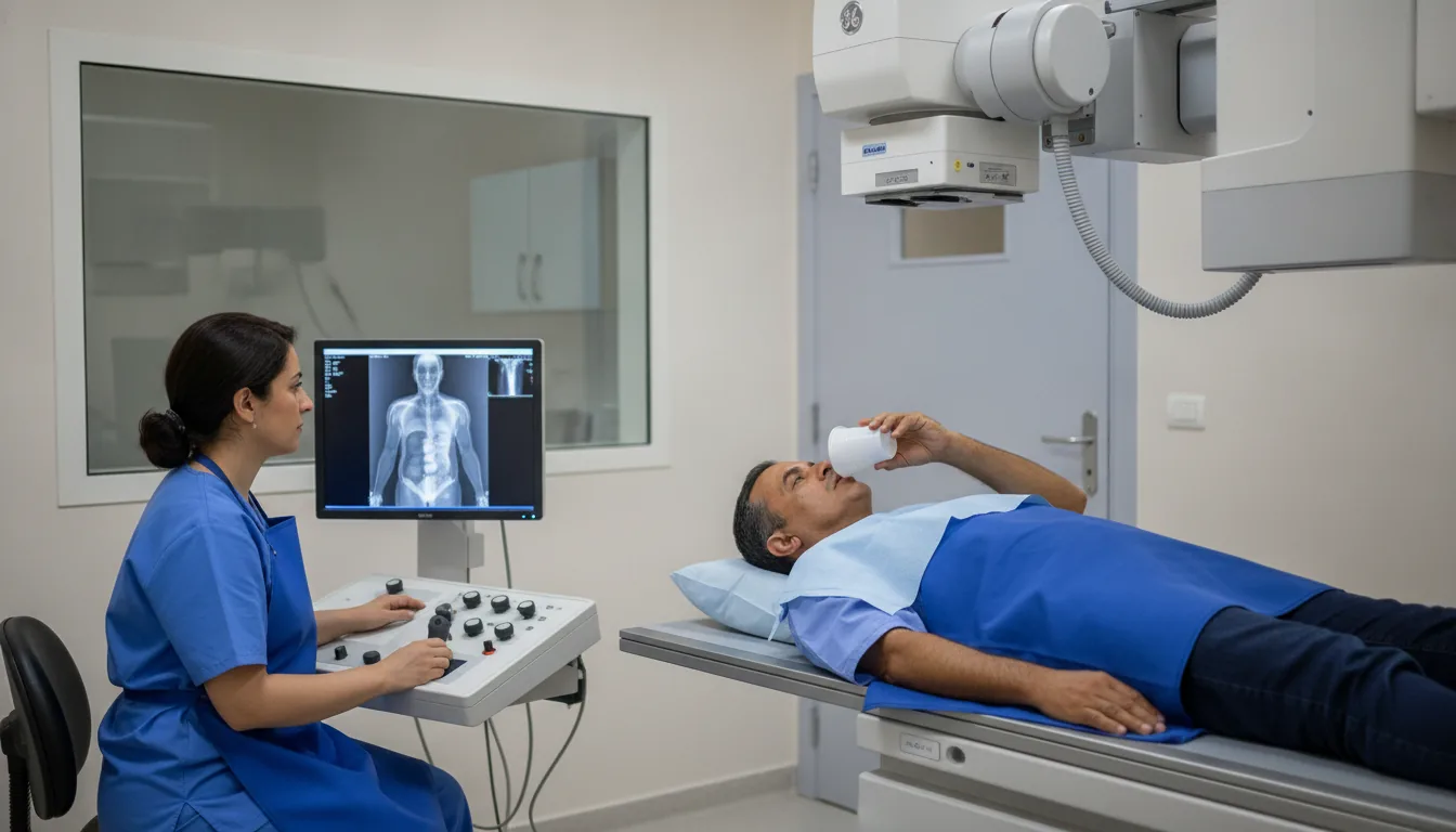
Venography (phlebography)
Venography in Tunis, Tunisia · Centre Urbain Nord
Venography in Tunis: X-ray of the veins of one or both limbs after injection of iodinated contrast, in specific cases.
What is it?
When is it indicated?
It provides an accurate map of the veins before a procedure, on medical advice.
When ultrasound cannot give an answer, it shows the inside of the veins directly.
It may show a narrowed, compressed or malformed vein.
Preparation
Procedure
You remove clothing and metal objects from the area being examined, in a changing cubicle
You lie on the X-ray table, sometimes slightly tilted
A small needle is placed in a vein in the foot or hand; a tourniquet may be tightened to direct the contrast
The contrast is injected and you may feel warmth in the limb
The radiologist follows the contrast agent live on a screen (fluoroscopy) and takes images at the right moments
After the examination, drink plenty of fluids and move the limb to flush out the contrast
Duration
About 30 minutes per limb
Results
The images are analysed by a radiologist, who writes a report. It is given to you with the images and sent to your doctor, who will explain the results and the next steps. You will be told on the day of the examination when the results will be ready.
Precautions and contraindications
Your doctor and the radiology team will inform you in detail before the examination. Feel free to ask them your questions.
This information is general and does not replace the advice of your doctor, who chooses the examination suited to your situation.
Other examinations — Conventional radiology
All examinationsRelated searches
- Venography Tunis
- Venography Tunisia
- Venography in Tunisia