
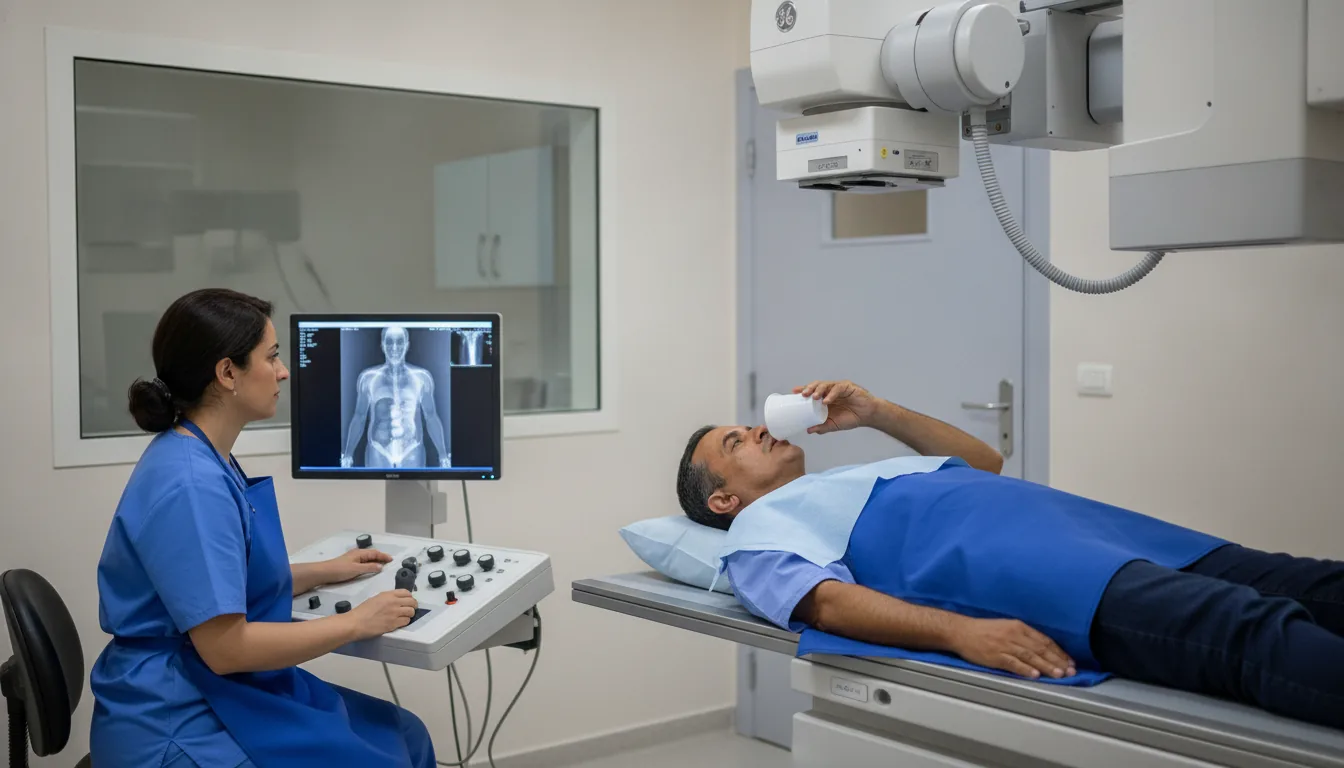
Chest X-ray (lungs, ribs)
Chest X-ray in Tunis, Tunisia · Centre Urbain Nord
Chest X-ray in Tunis: a quick examination of the lungs, heart and ribs for a cough, shortness of breath, fever or after an injury.
What is it?
When is it indicated?
The X-ray shows an infected area of the lung or fluid around the lung.
It helps look for a lung or heart cause and guides further tests.
The rib series looks for a rib fracture and checks for air or blood around the lung.
It is part of some check-ups before anaesthesia, or of medical files for a visa or work.
At the bedside, it monitors a lung condition or checks the position of a catheter or tube.
Preparation
Procedure
You remove clothing and metal objects from the area being examined, in a changing cubicle
You stand with your chest against the detector plate, shoulders rolled forward
You are asked to take a deep breath and hold it for a few seconds
Several images may be taken from different angles, called "views": from the front, from the side (lateral), sometimes at an oblique angle
At the bedside, the plate is slid behind your back and the unit is brought to you
After the examination, you can return to your usual activities straight away
Duration
About 5 to 10 minutes
Results
The images are analysed by a radiologist, who writes a report. It is given to you with the images and sent to your doctor, who will explain the results and the next steps. You will be told on the day of the examination when the results will be ready.
Precautions and contraindications
Your doctor and the radiology team will inform you in detail before the examination. Feel free to ask them your questions.
This information is general and does not replace the advice of your doctor, who chooses the examination suited to your situation.
Other examinations — Conventional radiology
Related searches
- Chest X-ray Tunis
- Chest X-ray Tunisia
- Chest X-ray in Tunisia