
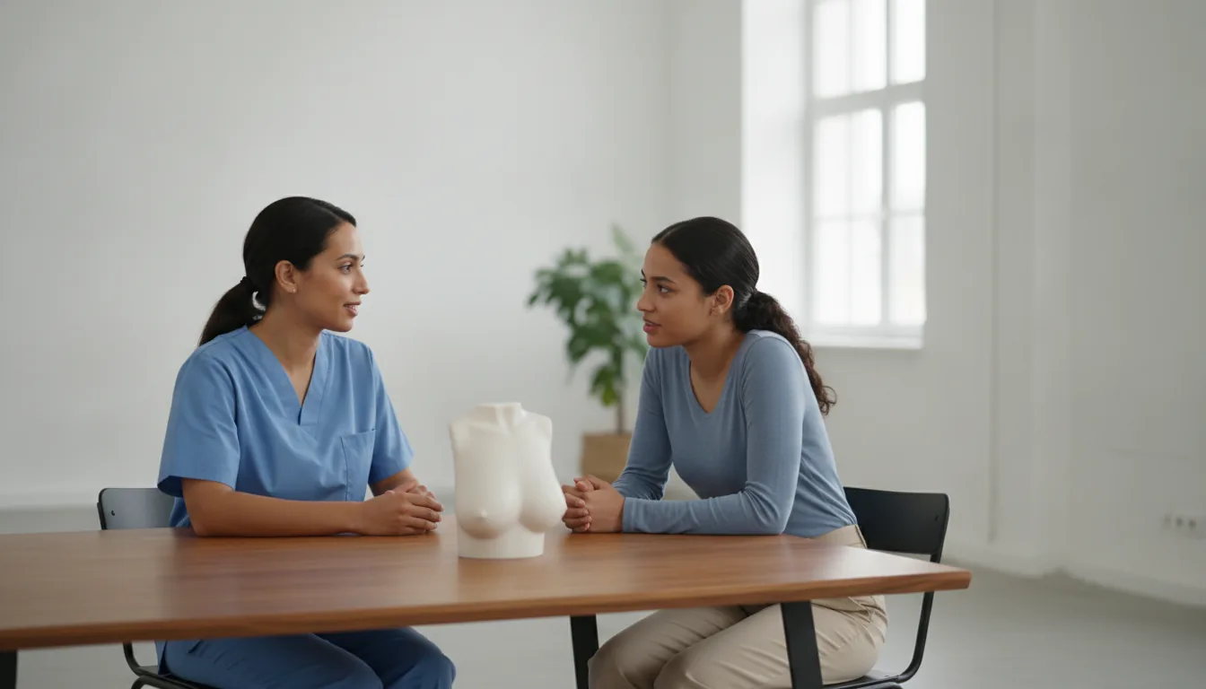
Removal of a benign breast lump
Surgeries/Gynecology-Obstetrics/Benign breast lump

Removal of a benign breast lump

Benign breast lump in Tunis, Tunisia · Centre Urbain Nord

Removal of a benign breast lump, such as a fibroadenoma, through a discreet incision that preserves the shape of the breast.

What is it?

Most breast lumps are benign, meaning they are not cancer. The most common in young women is the fibroadenoma, a firm, mobile, painless lump. Other benign lesions include papillomas (small growths in a milk duct, sometimes causing nipple discharge) and cysts. Many only need monitoring. Surgery is offered when the lump grows, becomes bothersome or painful, when a biopsy cannot give a clear answer, or at the patient's request after information. The surgeon removes the lesion through a short incision, often placed at the edge of the areola or in a natural fold, so that the scar is as discreet as possible. Before the operation, an ultrasound and, depending on age, a mammogram are carried out; a biopsy is often done to identify the nature of the lump. If the lesion cannot be felt, it is located by the radiologist on the day of surgery using ultrasound or mammography. At Clinique Pasteur Tunis, breast imaging and surgery are on the same site, which simplifies the patient's journey. The operation is a day-case procedure and the tissue removed is always analysed; the result is explained at the follow-up appointment.
Removal of a benign breast lump

When is it indicated?

Fibroadenoma that grows or exceeds a certain size

A fibroadenoma is benign, but if it grows or becomes large, it is better to remove it.

Lump that is bothersome, painful or a source of worry

Even if benign, a lump that causes daily discomfort or anxiety can be removed after discussion with the surgeon.

Inconclusive biopsy result

When the biopsy cannot confirm with certainty that the lesion is benign, removing it allows it to be analysed in full.

Nipple discharge from a single duct

Discharge, especially if it contains blood, may come from a papilloma. Removing the duct concerned identifies the cause.

High-risk lesion found on imaging

Some non-cancerous lesions are removed as a precaution because they need complete analysis.

Preparation

1Breast ultrasound, mammogram depending on age, and biopsy if indicated
2Localisation of the lesion by the radiologist on the morning of surgery if it cannot be felt
3Anaesthetic consultation
4Fasting for 6 hours before the operation
5Bring a comfortable non-wired bra for after the operation

Procedure

1

Admission as a day case

2

Short general anaesthetic, sometimes local anaesthetic

3

Discreet incision at the edge of the areola or in a natural fold

4

Removal of the lump with a thin margin of healthy tissue

5

Tissue sent for analysis

6

Closure with dissolvable stitches and dressing

7

Monitoring for a few hours, then discharge home

Duration

About 30 minutes to 1 hour

Aftercare & Recovery

Patients go home the same day. Pain is moderate and relieved by simple painkillers. Wearing a comfortable bra is advised for a few days. Activities resume within a few days, sport after two to three weeks. The scar fades gradually over the months. The analysis result is given at the follow-up appointment.

Risks and side effects

Haematoma or bruising of the breast, usually settling on its own
Wound infection, uncommon
Temporary change in sensation around the scar
Slight irregularity of the breast contour, particularly for large lumps
Another fibroadenoma may appear over time

These risks remain rare. Your surgeon will inform you in detail before the procedure.

Request a Quote

Contact us to schedule a consultation or receive a personalized quote.

Request a Quote +216 36 402 000

At a glance

Duration: About 30 minutes to 1 hour
Preparation: Required
Dedicated specialist team

Related searches

  • Benign breast lump Tunis
  • Benign breast lump Tunisia
  • Benign breast lump in Tunisia
Request a quote