
Pelvic MRI (uterus, ovaries, rectum)
Pelvic MRI in Tunis, Tunisia · Centre Urbain Nord
Pelvic MRI in Tunis: examines the uterus, ovaries, bladder and rectum (fibroids, endometriosis, cysts, assessment of certain cancers).
What is it?
When is it indicated?
It counts and locates fibroids to help choose between monitoring, medicines, surgery or embolisation.
It looks for endometriosis lesions in the pelvis and their extent, before a treatment decision.
It clarifies the nature of a mass that is poorly characterised on ultrasound.
It measures the local extent of the tumour, which guides treatment.
It looks for a cause in the uterus, ovaries or nearby tissues.
Preparation
Procedure
You change in a cubicle and put your metal objects in a locker
A drip is placed in your arm if an injection is planned; a medicine to calm bowel movements may be injected
For certain indications, a gel is placed in the vagina or rectum with a small tube, with your consent
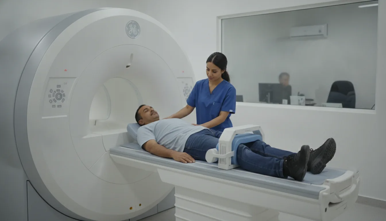
You lie on your back, with a coil placed on your lower abdomen
The scanner makes loud, repetitive noises while the images are being taken: headphones or earplugs are provided
You need to keep still during each series of images so that they are sharp
You stay in contact with the team through a microphone and a call bell that you hold in your hand
Duration
About 30 to 45 minutes
Results
The images are analysed by a radiologist, who writes a report. The report is given to you with the images and sent to your doctor, who will explain the results and the next steps. You will be told when to expect it on the day of the examination.
Precautions and contraindications
Your doctor and the radiology team will inform you in detail before the examination. Feel free to ask them your questions.
This information is general and does not replace the advice of your doctor, who chooses the examination suited to your situation.
Related searches
- Pelvic MRI Tunis
- Pelvic MRI Tunisia
- Pelvic MRI in Tunisia