
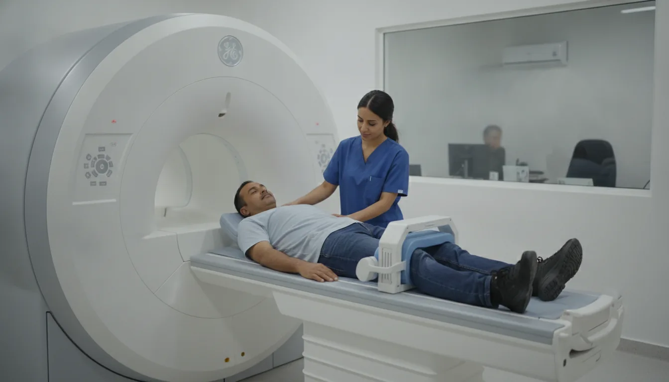
MRI of the bony pelvis and hips
Pelvis and hip MRI in Tunis, Tunisia · Centre Urbain Nord
Pelvis MRI in Tunis: pelvic bones, hips, sacroiliac joints and nearby muscles, for hip or buttock pain.
What is it?
When is it indicated?
It can show early damage to the head of the femur or a fracture that is invisible on X-ray.
It looks for inflammation of the sacroiliac joints, useful in diagnosing certain rheumatic diseases.
Especially in athletes, it identifies a muscle tear or a tendon problem.
It characterises a bone abnormality found on another test.
Preparation
Procedure
You change in a cubicle and put your metal objects in a locker
You lie on your back, with a coil placed on your pelvis; your feet may be held in place to stop them from turning
The scanner makes loud, repetitive noises while the images are being taken: headphones or earplugs are provided
You need to keep still during each series of images so that they are sharp
If an injection is needed, the contrast agent (gadolinium) is given through a small drip in your arm during the examination
You stay in contact with the team through a microphone and a call bell that you hold in your hand
After the examination, you can return to your usual activities straight away
Duration
About 20 to 30 minutes
Results
The images are analysed by a radiologist, who writes a report. The report is given to you with the images and sent to your doctor, who will explain the results and the next steps. You will be told when to expect it on the day of the examination.
Precautions and contraindications
Your doctor and the radiology team will inform you in detail before the examination. Feel free to ask them your questions.
This information is general and does not replace the advice of your doctor, who chooses the examination suited to your situation.
Related searches
- Pelvis and hip MRI Tunis
- Pelvis and hip MRI Tunisia
- Pelvis and hip MRI in Tunisia