
Pituitary MRI (MRI of the pituitary gland)
Centres/Radiology/MRI/Pituitary MRI

Pituitary MRI (MRI of the pituitary gland)

Pituitary MRI in Tunis, Tunisia · Centre Urbain Nord

Pituitary MRI in Tunis: a targeted scan of the small gland at the base of the brain, requested for a hormonal disorder or a vision problem.

What is it?

The pituitary gland is a small gland located at the base of the brain, just behind the eyes. It controls many other glands (thyroid, adrenal glands, ovaries, testicles) and milk production. A pituitary MRI is a brain MRI focused on this region, with very thin slices. It is most often requested by an endocrinologist when a blood test shows a hormonal imbalance, or by an ophthalmologist when certain areas of vision are reduced. An injection of contrast agent (gadolinium) is usual: the images taken during the injection make it possible to detect very small lesions, most often benign (adenomas).

When is it indicated?

Excess prolactin or other hormonal disorder

High prolactin, irregular periods or milk discharge outside breastfeeding may lead to a search for a small pituitary adenoma.

Narrowing of the field of vision

The pituitary gland is close to the optic nerves: a lesion can reduce vision at the sides.

Follow-up of a known adenoma

MRI checks the size of the lesion during treatment or after surgery.

Delayed growth or puberty in children

On the endocrinologist's advice, it examines the gland that controls growth and puberty.

Preparation

1Remove jewellery, watch, piercings, hair clips, bank cards and any metal object before entering the scanner room
2Tell us in advance about any pacemaker or defibrillator, implant (cochlear implant, neurostimulator, pump), surgical clip, heart valve, prosthesis or metal fragment: some are MRI-compatible, others are not
3Tell us if you are pregnant or think you might be
4Let us know if you are afraid of enclosed spaces (claustrophobia): the team will support you and your doctor can prescribe a mild sedative
5If an injection is planned, tell us about any kidney disease, allergy or past reaction to a contrast agent; a recent blood test (creatinine) may be requested
6Bring your hormone test results and your referral
7No fasting is usually needed

Procedure

1

You change in a cubicle and put your metal objects in a locker

2

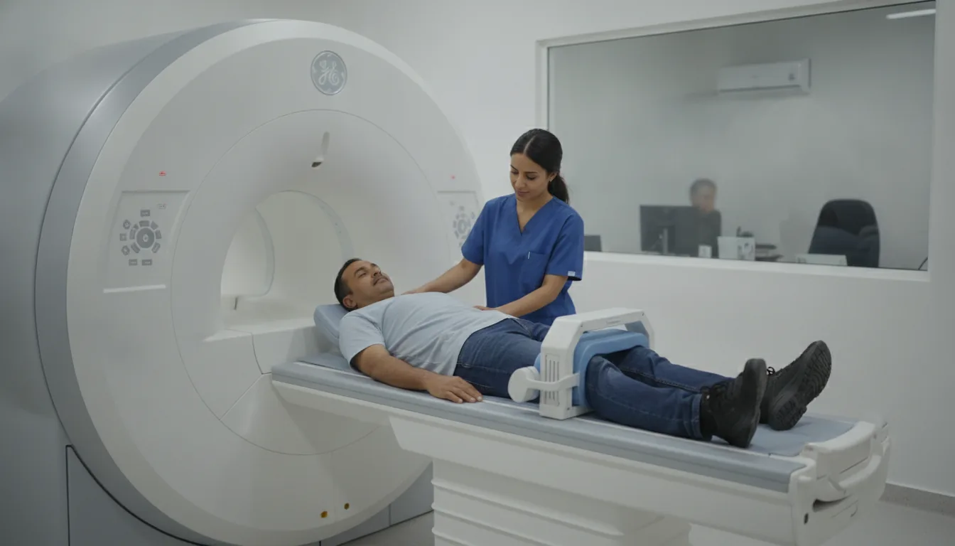
You lie on your back, with your head in a helmet-shaped coil

3

A drip is placed in your arm before the images start

4

The scanner makes loud, repetitive noises while the images are being taken: headphones or earplugs are provided

5

The contrast agent is injected during a series of rapid images: it is important not to move at that moment

6

You stay in contact with the team through a microphone and a call bell that you hold in your hand

7

After the examination, you can return to your usual activities straight away

Duration

About 20 to 30 minutes

Results

The images are analysed by a radiologist, who writes a report. The report is given to you with the images and sent to your doctor, who will explain the results and the next steps. You will be told when to expect it on the day of the examination.

Precautions and contraindications

No X-rays: MRI does not use ionising radiation
Not possible with a pacemaker, defibrillator or implant that is not MRI-compatible, or with a metal foreign body in the eye
Allergic reaction to gadolinium: rare, and usually mild (redness, itching)
In case of severe kidney failure, the gadolinium injection is avoided or adjusted by the doctor
During pregnancy, MRI is only performed if it is really useful, and usually without an injection
You may feel enclosed inside the tunnel; the examination can be stopped at any time

Your doctor and the radiology team will inform you in detail before the examination. Feel free to ask them your questions.

This information is general and does not replace the advice of your doctor, who chooses the examination suited to your situation.

Related searches

  • Pituitary MRI Tunis
  • Pituitary MRI Tunisia
  • Pituitary MRI in Tunisia
Request a quoteBook an appointment