
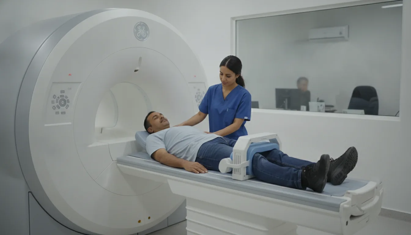
Knee MRI
Knee MRI in Tunis, Tunisia · Centre Urbain Nord
Knee MRI in Tunis: menisci, cruciate ligaments, cartilage and tendons, after a sprain, locking or knee pain.
What is it?
When is it indicated?
It looks for a torn cruciate ligament, a meniscus tear or a bone bruise.
It often finds the mechanical cause: a fragment of meniscus or a cartilage injury.
It assesses cartilage wear, tendinitis or a kneecap problem.
It helps prepare the operation, then check the graft or repair.
Preparation
Procedure
You change in a cubicle and put your metal objects in a locker
You lie on your back, feet first; your knee is placed in a tube-shaped coil and supported with cushions
Most of the time, your head and upper body stay outside the tunnel
The scanner makes loud, repetitive noises while the images are being taken: headphones or earplugs are provided
You need to keep still during each series of images so that they are sharp
You stay in contact with the team through a microphone and a call bell that you hold in your hand
After the examination, you can return to your usual activities straight away
Duration
About 15 to 30 minutes
Results
The images are analysed by a radiologist, who writes a report. The report is given to you with the images and sent to your doctor, who will explain the results and the next steps. You will be told when to expect it on the day of the examination.
Precautions and contraindications
Your doctor and the radiology team will inform you in detail before the examination. Feel free to ask them your questions.
This information is general and does not replace the advice of your doctor, who chooses the examination suited to your situation.
Other examinations — MRI
All examinationsRelated searches
- Knee MRI Tunis
- Knee MRI Tunisia
- Knee MRI in Tunisia