
Elbow MRI

Elbow MRI

Elbow MRI in Tunis, Tunisia · Centre Urbain Nord

Elbow MRI in Tunis: tendons, ligaments, cartilage and nerves of the elbow, for persistent pain, injury or tingling.

What is it?

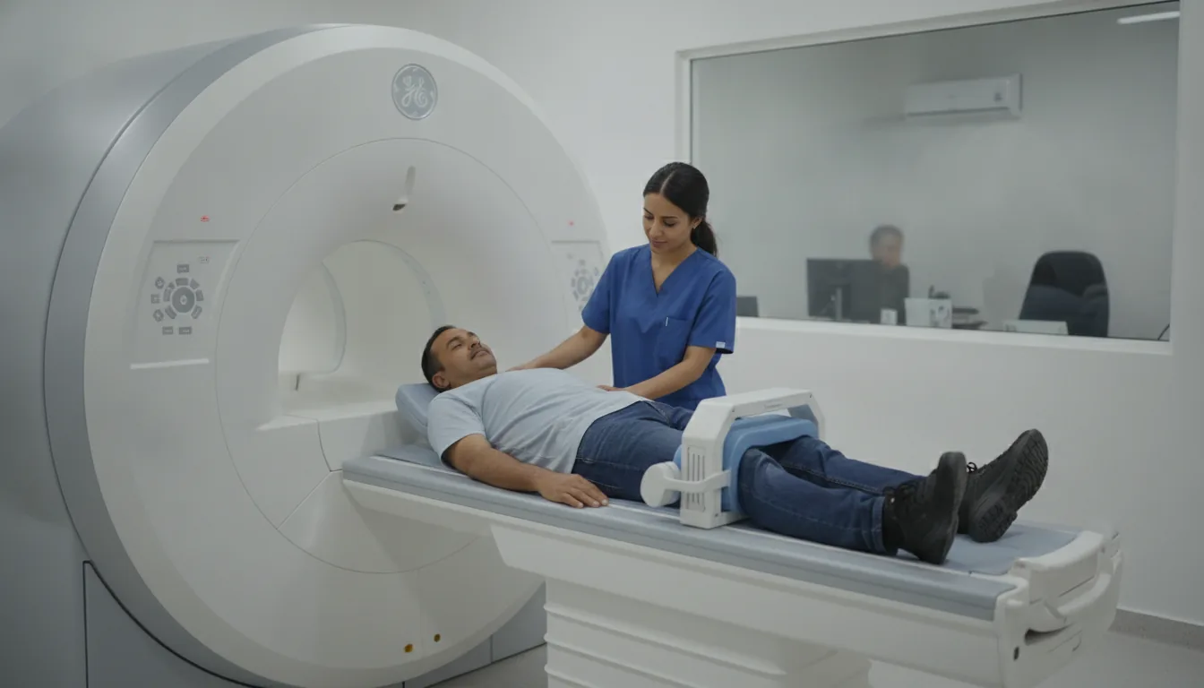
An elbow MRI examines the tendons attached around the elbow, the ligaments that stabilise it, the cartilage, the bones and the nerves running through it (including the ulnar nerve, which gives sensation to the last fingers). It is requested for elbow pain that does not respond to treatment (for example stubborn lateral epicondylitis, or "tennis elbow"), after an injury, or in case of locking or tingling in the last fingers. It is most often performed without an injection. Depending on the scanner and your build, your arm is placed by your side or above your head.

When is it indicated?

Epicondylitis or tendinitis that does not respond to treatment

It assesses the condition of the tendon and looks for a tear.

Elbow injury

It looks for damage to a ligament or cartilage, or a fracture invisible on X-ray.

Locking or clicking of the elbow

It can show a loose fragment of cartilage or bone in the joint.

Tingling in the last fingers

It examines the ulnar nerve where it passes the elbow, alongside the electromyogram (EMG).

Preparation

1Remove jewellery, watch, piercings, hair clips, bank cards and any metal object before entering the scanner room
2Tell us in advance about any pacemaker or defibrillator, implant (cochlear implant, neurostimulator, pump), surgical clip, heart valve, prosthesis or metal fragment: some are MRI-compatible, others are not
3Tell us about any orthopaedic hardware in the elbow
4Tell us if you are pregnant or think you might be
5Let us know if you are afraid of enclosed spaces (claustrophobia): the team will support you and your doctor can prescribe a mild sedative
6Bring your elbow X-rays and ultrasounds

Procedure

1

You change in a cubicle and put your metal objects in a locker

2

You lie down with your elbow placed in a coil, your arm by your side or above your head, whichever position is more comfortable

3

The scanner makes loud, repetitive noises while the images are being taken: headphones or earplugs are provided

4

You need to keep still during each series of images so that they are sharp

5

You stay in contact with the team through a microphone and a call bell that you hold in your hand

6

After the examination, you can return to your usual activities straight away

Duration

About 20 to 30 minutes

Results

The images are analysed by a radiologist, who writes a report. The report is given to you with the images and sent to your doctor, who will explain the results and the next steps. You will be told when to expect it on the day of the examination.

Precautions and contraindications

No X-rays: MRI does not use ionising radiation
Not possible with a pacemaker, defibrillator or implant that is not MRI-compatible, or with a metal foreign body in the eye
An injection is rarely needed; if it is, allergic reaction to gadolinium is rare
During pregnancy, MRI is only performed if it is really useful, and usually without an injection
You may feel enclosed inside the tunnel; the examination can be stopped at any time

Your doctor and the radiology team will inform you in detail before the examination. Feel free to ask them your questions.

This information is general and does not replace the advice of your doctor, who chooses the examination suited to your situation.

Related searches

  • Elbow MRI Tunis
  • Elbow MRI Tunisia
  • Elbow MRI in Tunisia
Request a quoteBook an appointment