
MRCP (MR cholangiopancreatography)

MRCP (MR cholangiopancreatography)

MRCP in Tunis, Tunisia · Centre Urbain Nord

MRCP in Tunis: MRI of the bile ducts and pancreatic duct to look for a stone or narrowing, without endoscopy or X-rays.

What is it?

MRCP (MR cholangiopancreatography) is an MRI that shows the ducts carrying bile from the liver to the intestine (bile ducts, including the common bile duct) as well as the pancreatic duct. It relies on MRI's ability to show fluids in white: the bile then appears as if drawn on a map, without injecting anything into the ducts and without endoscopy. It can look for a stone stuck in the common bile duct, or a narrowing or widening of the ducts. It is usually performed without a gadolinium injection and is often combined with an MRI of the liver and pancreas. Fasting is essential so that the gallbladder is well filled and the stomach empty.

When is it indicated?

Suspected stone in the common bile duct

After pain, jaundice or abnormal liver tests, it looks for a stone that has passed from the gallbladder into the bile ducts.

Unexplained jaundice

It shows where and why bile is no longer flowing normally.

Pancreatitis

It looks for a cause (stone, abnormality of the pancreatic duct) and for complications.

Assessment before gallbladder removal or biliary endoscopy

It helps the doctor know whether a procedure on the bile ducts is needed.

Preparation

1Remove jewellery, watch, piercings, hair clips, bank cards and any metal object before entering the scanner room
2Tell us in advance about any pacemaker or defibrillator, implant (cochlear implant, neurostimulator, pump), surgical clip, heart valve, prosthesis or metal fragment: some are MRI-compatible, others are not
3Fast for at least 4 to 6 hours (no meals, no sugary drinks, no smoking); only small sips of water are allowed, unless told otherwise
4Tell us if you are pregnant or think you might be
5Let us know if you are afraid of enclosed spaces (claustrophobia): the team will support you and your doctor can prescribe a mild sedative
6Bring your blood test results (liver tests) and your recent ultrasound

Procedure

1

You change in a cubicle and put your metal objects in a locker

2

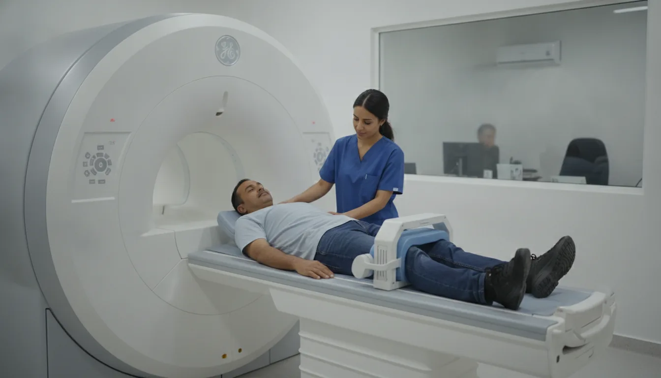
You lie on your back, with a coil placed on your abdomen

3

A drink or medicine may be given to reduce the signal from the stomach and intestine

4

The series of images are taken while you hold your breath for a few seconds or breathe calmly and regularly

5

The scanner makes loud, repetitive noises while the images are being taken: headphones or earplugs are provided

6

You stay in contact with the team through a microphone and a call bell that you hold in your hand

7

After the examination, you can return to your usual activities straight away

Duration

About 20 to 30 minutes

Results

The images are analysed by a radiologist, who writes a report. The report is given to you with the images and sent to your doctor, who will explain the results and the next steps. You will be told when to expect it on the day of the examination.

Precautions and contraindications

No X-rays: MRI does not use ionising radiation
Not possible with a pacemaker, defibrillator or implant that is not MRI-compatible, or with a metal foreign body in the eye
Usually performed without an injection; if one is added, allergic reaction to gadolinium is rare
During pregnancy, MRI is only performed if it is really useful, and usually without an injection
You may feel enclosed inside the tunnel; the examination can be stopped at any time

Your doctor and the radiology team will inform you in detail before the examination. Feel free to ask them your questions.

This information is general and does not replace the advice of your doctor, who chooses the examination suited to your situation.

Related searches

  • MRCP Tunis
  • MRCP Tunisia
  • MRCP in Tunisia
Request a quoteBook an appointment