
FFR measurement and intracoronary imaging (IVUS, OCT)

FFR measurement and intracoronary imaging (IVUS, OCT)

FFR, IVUS and OCT in Tunis, Tunisia · Centre Urbain Nord

Tests performed during coronary angiography to measure the real significance of a narrowing and to see inside the artery.

What is it?

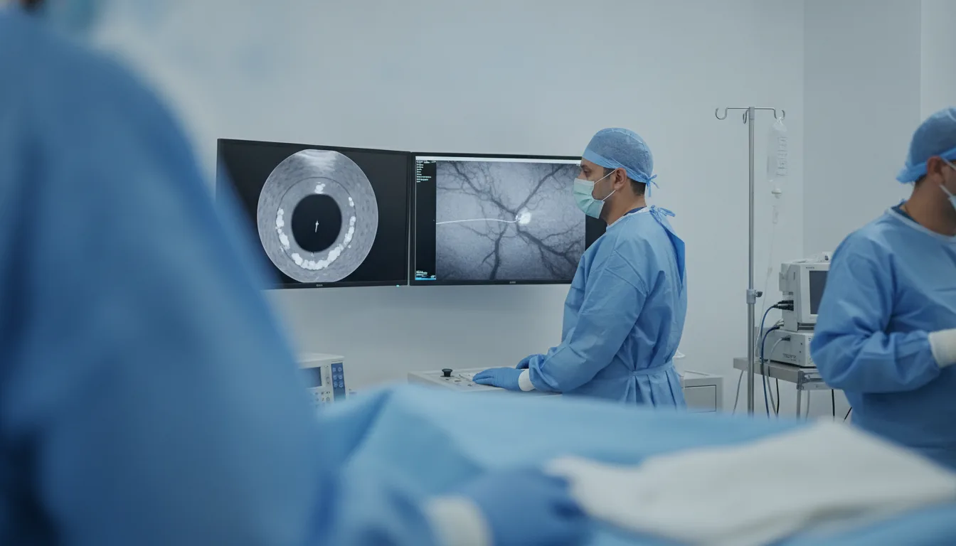
Coronary angiography shows the outline of the coronary arteries, but it does not always tell whether a narrowing truly restricts blood flow, nor what the artery wall looks like. Two types of complementary tests, performed during the angiogram through the same access, answer these questions. FFR (fractional flow reserve) measurement uses a very fine guidewire fitted with a pressure sensor, placed beyond the narrowing. The pressure beyond and before the narrowing is compared, usually after injection of a medicine that increases coronary blood flow; resting indices measured without this medicine (such as iFR) are also available. A low value means the narrowing limits blood flow and should be treated; a normal value often avoids an unnecessary stent. Intracoronary imaging shows the inside of the artery: IVUS (intravascular ultrasound) uses sound waves, while OCT (optical coherence tomography) uses light and gives a very detailed image. They make it possible to measure the artery diameter, analyse the plaque and calcium, choose the right stent size and check after implantation that the stent is fully open and well apposed to the wall. At Clinique Pasteur Tunis, these measurements are performed in a latest-generation catheterisation laboratory whenever the cardiologist considers that they will provide useful information for the decision or for the quality of the angioplasty.
FFR measurement and intracoronary imaging (IVUS, OCT)

When is it indicated?

Intermediate narrowing of uncertain significance

When the image cannot show whether the narrowing really restricts blood flow, FFR settles the question and avoids placing a stent for no reason.

Multivessel disease

Measuring each narrowing helps decide which ones to treat and choose between angioplasty and bypass surgery.

Left main or complex lesion angioplasty

Intracoronary imaging measures the artery precisely and checks that the stents are well deployed, which improves the long-term result.

Heavily calcified plaques

Seeing the thickness and extent of the calcium helps decide whether it needs to be treated before a stent is placed.

Assessment of a stent that has narrowed or blocked

Imaging reveals the cause of the problem (under-expanded stent, new plaque…) and guides the appropriate treatment.

Preparation

1Same preparation as for coronary angiography: blood tests including kidney function
2Report any allergy, especially to iodine
3Report severe asthma, which may favour a measurement without the flow-increasing medicine
4Adjustment of anticoagulant and diabetes medicines as instructed
5Fasting for a few hours before the test

Procedure

1

Coronary angiography through the wrist or groin, under local anaesthesia

2

Injection of an anticoagulant to allow the instruments to be passed safely

3

For FFR: the pressure-sensor guidewire is passed beyond the narrowing and the measurement taken

4

For IVUS or OCT: a small imaging probe is passed and records the inside of the artery in a few seconds

5

Immediate analysis of the results and decision whether or not to treat the narrowing

Duration

10 to 30 minutes longer than a coronary angiogram

Results

Results are available immediately and guide the next step: stent treatment in the same session, monitoring with medical treatment, or discussion of bypass surgery. Recovery is that of the angiogram or angioplasty performed, usually with discharge the same day or the next day.

Risks and side effects

Same risks as coronary angiography, low overall
Brief feeling of warmth, chest tightness or breathlessness when the FFR medicine is injected
Spasm of the artery in contact with the probe, treated immediately with medication
Rare coronary complication, managed on the spot

These risks remain rare. Your physician will inform you in detail before the examination.

Request a quote

Request your quote online or call us on the number below:

Request a Quote +216 36 402 000

At a glance

Duration: 10 to 30 minutes longer than a coronary angiogram
Preparation: Required

Related searches

  • FFR, IVUS and OCT Tunis
  • FFR, IVUS and OCT Tunisia
  • FFR, IVUS and OCT in Tunisia
Request a quoteBook an appointment