
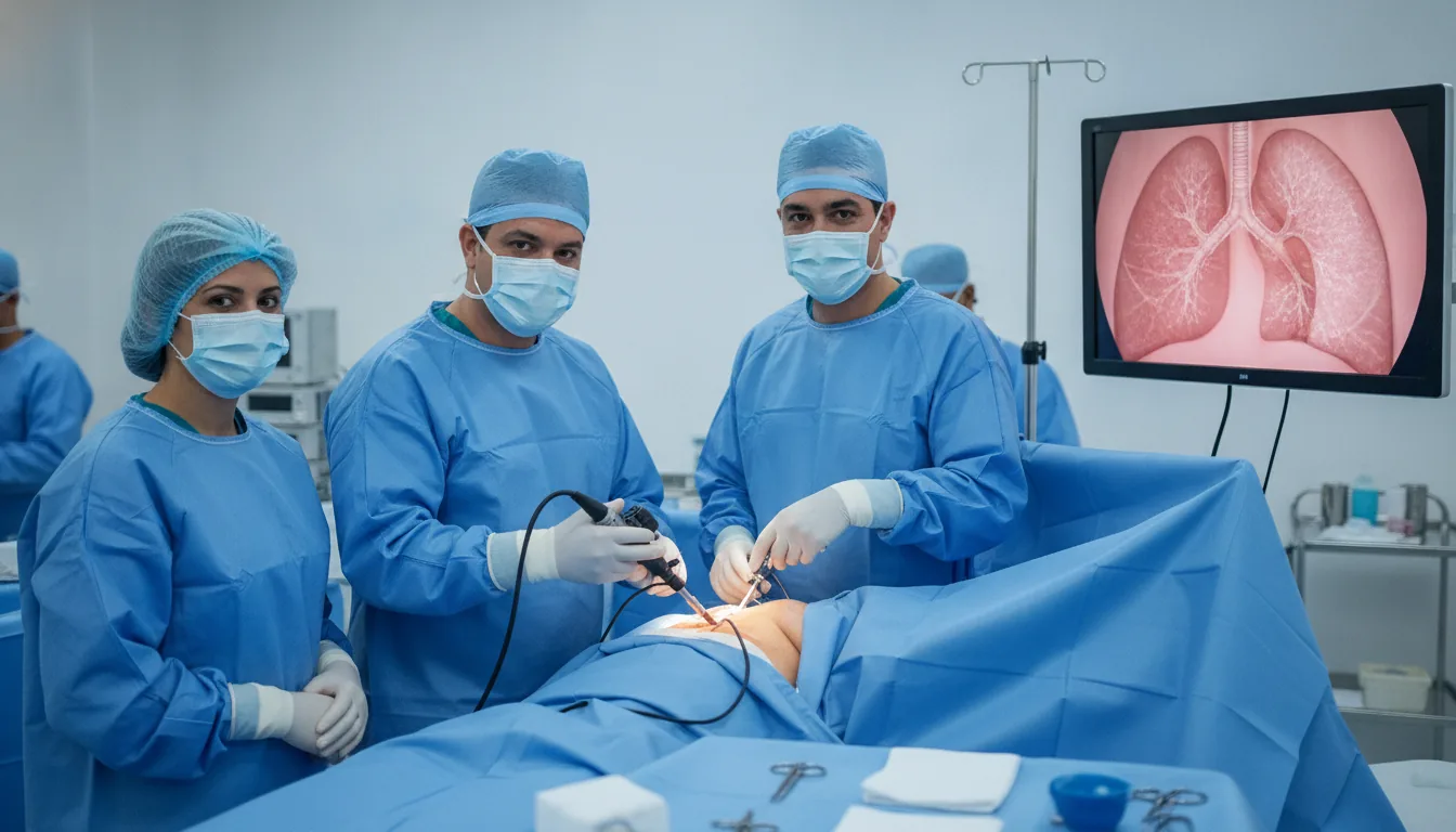
Thoracoscopy (pleural biopsy, pleural effusion, pneumothorax)
Thoracoscopy in Tunis, Tunisia · Centre Urbain Nord
Examination and treatment of the pleura using a camera passed between two ribs: biopsy, drainage of an effusion or treatment of a pneumothorax.
What is it?
When is it indicated?
When fluid builds up around the lung with no explanation, samples of the pleura taken under direct vision help find the cause.
An infection of the pleura can form pockets of pus; thoracoscopy cleans them out so that the lung can expand again.
When the lung has collapsed several times, the procedure treats the blisters responsible and seals the pleura to prevent further episodes.
If air keeps leaking despite drainage, thoracoscopy allows the leak to be closed.
When fluid comes back after every tap, the pleura can be sealed to avoid repeated drainage.
Preparation
Procedure
General anaesthesia
Patient positioned on their side
One to three small incisions between the ribs
Introduction of the camera and examination of the pleural cavity
Appropriate procedure: biopsies, drainage of the effusion, treatment of blisters, pleurodesis
Placement of a chest drain and closure of the incisions
Monitoring in the recovery room, then in the ward
Duration
About 30 minutes to 1 hour 30
Aftercare & Recovery
The hospital stay generally lasts 2 to 5 days, until the drain is removed once the lung is fully expanded. Pain between the ribs is common and well relieved by painkillers. Chest physiotherapy helps recovery. Activities are resumed gradually over 2 to 4 weeks; flying and diving should be avoided for some time after a pneumothorax, as advised by the surgeon. Biopsy results are discussed at a follow-up visit.
Risks and side effects
These risks remain rare. Your surgeon will inform you in detail before the procedure.
Request a Quote
Contact us to schedule a consultation or receive a personalized quote.
Request a Quote +216 36 402 000At a glance
Related searches
- Thoracoscopy Tunis
- Thoracoscopy Tunisia
- Thoracoscopy in Tunisia