
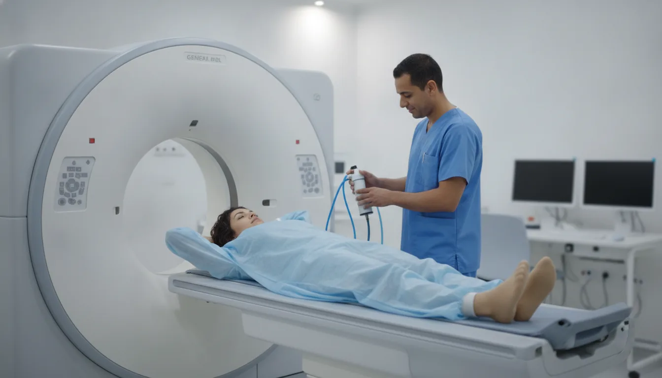
Dental CT and cone beam CT (3D imaging of the jaws)
Dental CT and cone beam in Tunis, Tunisia · Centre Urbain Nord
Dental CT and cone beam CT in Tunis: 3D imaging of the jaws before a dental implant, for an impacted tooth or a cyst, without injection.
What is it?
When is it indicated?
It measures the available bone and locates the jaw nerve and sinuses so the implant can be placed safely.
It precisely locates a tooth that has stayed in the bone (often a wisdom tooth) and its relationship to the nerve.
It shows the size and extent of the lesion before treatment.
It looks for a bone infection or sinus involvement of dental origin.
Preparation
Procedure
For dental CT, you lie on your back; for cone beam CT, you usually sit or stand with your head held still
A small bite block may be placed between your teeth to keep the two jaws apart
You need to keep still, not swallow and not move your tongue during the acquisition
Image acquisition is quick: from a few seconds to a few minutes
You can leave straight away, with no special instructions
Duration
About 5 to 15 minutes in total
Results
The images are analysed by a radiologist, who writes a report. The report is given to you with the images and sent to your doctor, who will explain the results and the next steps. You will be told when to expect it on the day of the examination.
Precautions and contraindications
Your doctor and the radiology team will inform you in detail before the examination. Feel free to ask them your questions.
This information is general and does not replace the advice of your doctor, who chooses the examination suited to your situation.
Other examinations — CT scan
All examinationsRelated searches
- Dental CT and cone beam Tunis
- Dental CT and cone beam Tunisia
- Dental CT and cone beam in Tunisia