
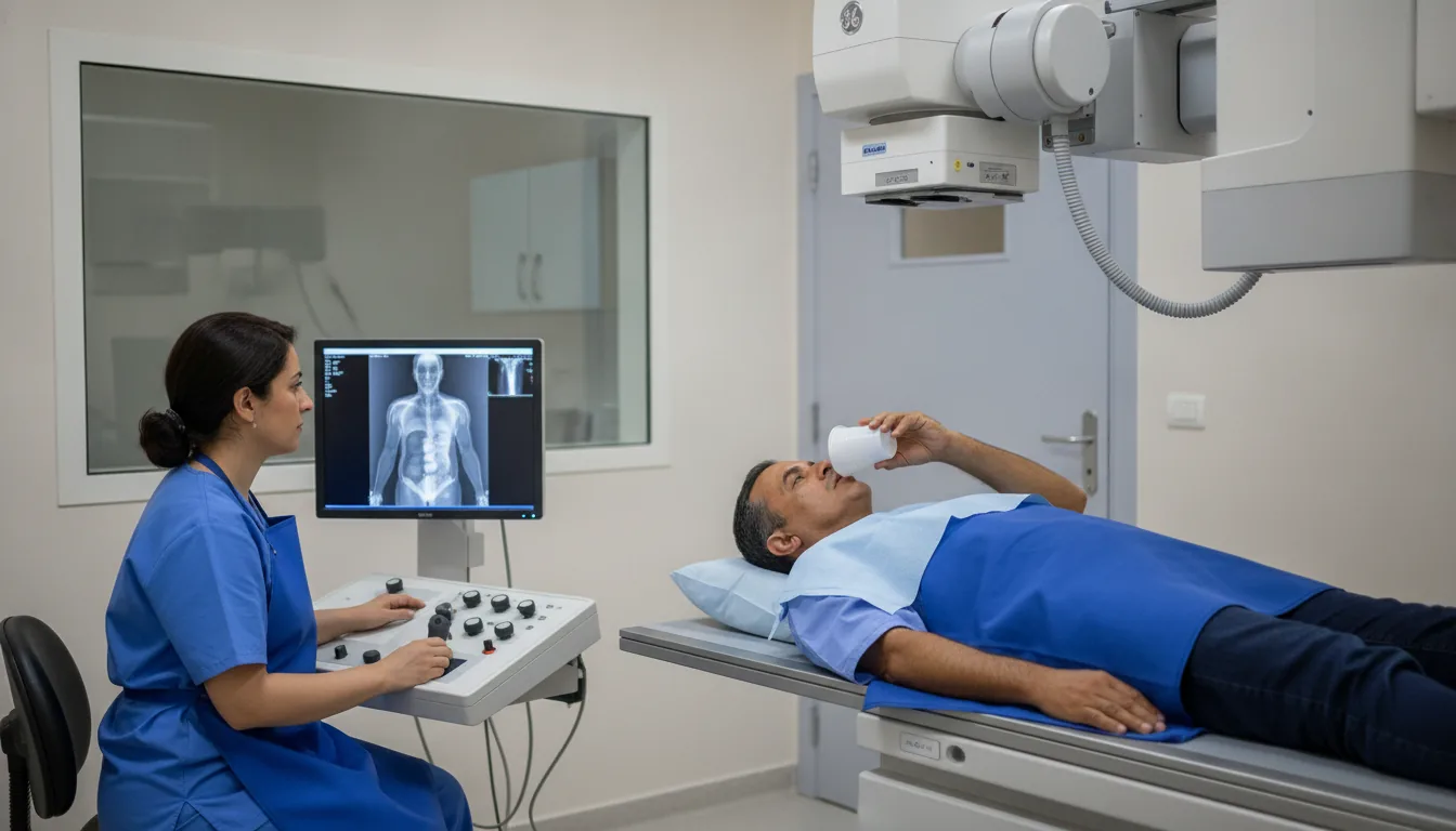
Long-length X-ray (whole spine or lower limbs)
Long-length X-ray in Tunis, Tunisia · Centre Urbain Nord
Long-length X-ray in Tunis: a standing full-length image of the whole spine or legs (scoliosis, leg length difference, knee alignment).
What is it?
When is it indicated?
It measures the angle of the spinal curve and helps monitor its progression, especially during growth.
It measures the difference in length precisely, for example to fit a shoe insert.
Before a knee replacement or alignment correction, it shows whether the legs are bowed or knock-kneed.
It assesses the overall balance of the spine and pelvis.
Preparation
Procedure
You remove clothing and metal objects from the area being examined, in a changing cubicle
You stand still in front of a large plate, feet positioned as instructed by the radiographer
A graduated ruler may be placed next to you to allow measurements
During each image you need to keep still for a few seconds; you may be asked to hold your breath
For the spine, a second image may be taken from the side
After the examination, you can return to your usual activities straight away
Duration
About 10 to 15 minutes
Results
The images are analysed by a radiologist, who writes a report. It is given to you with the images and sent to your doctor, who will explain the results and the next steps. You will be told on the day of the examination when the results will be ready.
Precautions and contraindications
Your doctor and the radiology team will inform you in detail before the examination. Feel free to ask them your questions.
This information is general and does not replace the advice of your doctor, who chooses the examination suited to your situation.
Other examinations — Conventional radiology
Related searches
- Long-length X-ray Tunis
- Long-length X-ray Tunisia
- Long-length X-ray in Tunisia