
Skull, face and sinus X-rays
Skull, face and sinus X-rays in Tunis, Tunisia · Centre Urbain Nord
Skull, sinus, nasal bone, orbit, ear and TMJ X-rays in Tunis, and lateral nasopharynx X-ray for children (adenoids).
What is it?
When is it indicated?
Images of the nasal bones or face look for a fracture.
It may show a filled sinus; a CT scan is often preferred if surgery is being considered.
TMJ images compare the position of the joint with the mouth open and closed.
The lateral nasopharynx view assesses the size of the adenoids before the ENT specialist decides.
Preparation
Procedure
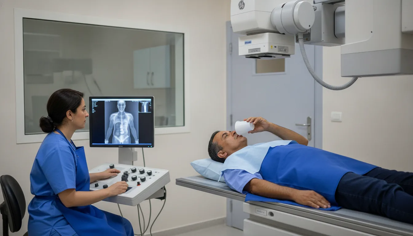
You sit or stand with your head resting against the plate; small supports help keep it in the right position
Several images may be taken from different angles, called "views": from the front, from the side (lateral), sometimes at an oblique angle
For the TMJ, one image is taken with the mouth open and another with the mouth closed
During each image you need to keep still for a few seconds; you may be asked to hold your breath
The radiographer checks that the images are sharp before you get dressed
After the examination, you can return to your usual activities straight away
Duration
About 5 to 15 minutes
Results
The images are analysed by a radiologist, who writes a report. It is given to you with the images and sent to your doctor, who will explain the results and the next steps. You will be told on the day of the examination when the results will be ready.
Precautions and contraindications
Your doctor and the radiology team will inform you in detail before the examination. Feel free to ask them your questions.
This information is general and does not replace the advice of your doctor, who chooses the examination suited to your situation.
Other examinations — Conventional radiology
Related searches
- Skull, face and sinus X-rays Tunis
- Skull, face and sinus X-rays Tunisia
- Skull, face and sinus X-rays in Tunisia