
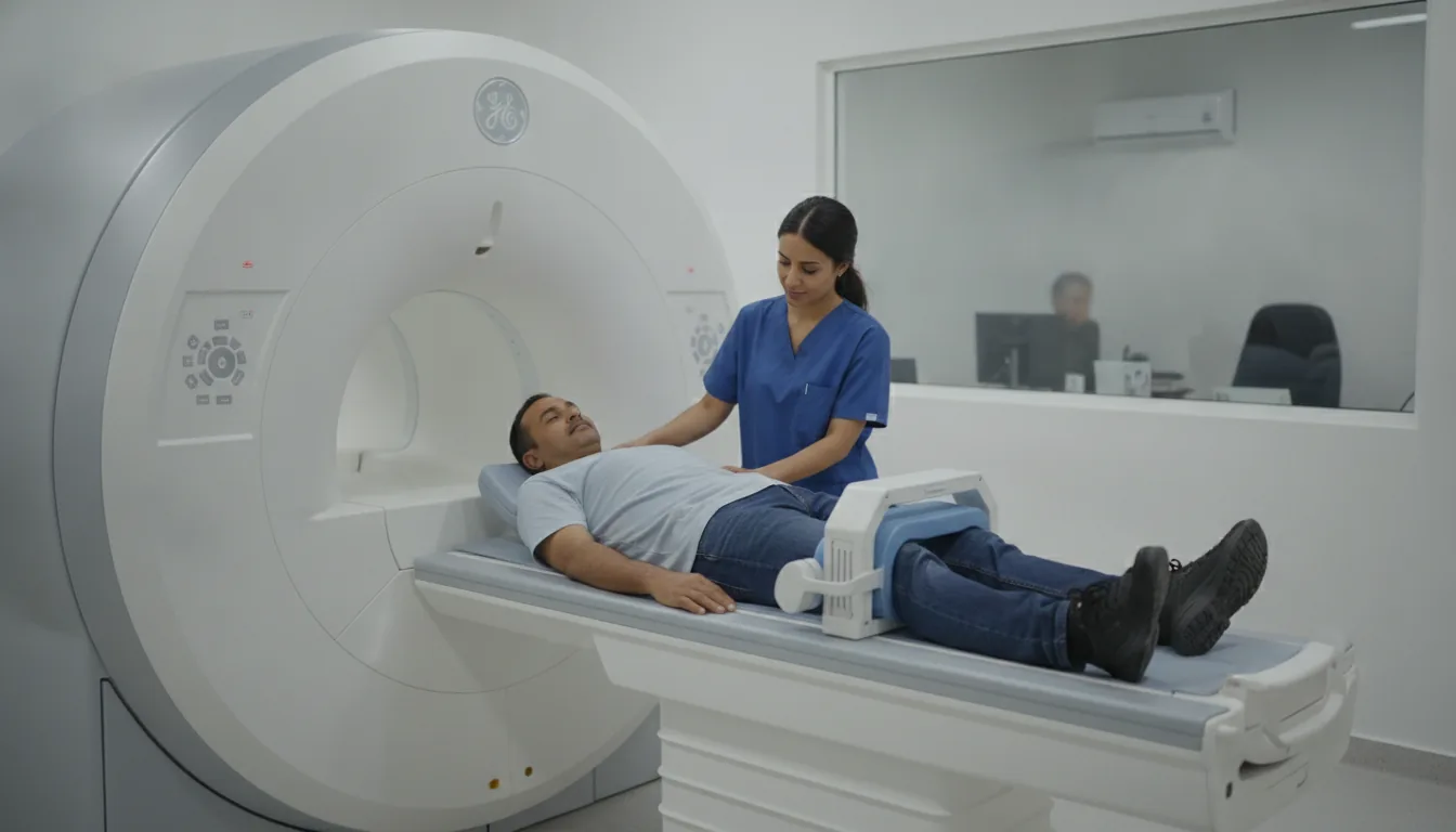
Spine MRI (spinal cord MRI)

Spine MRI (spinal cord MRI)

Spine MRI in Tunis, Tunisia · Centre Urbain Nord

Cervical, thoracic or lumbar spine MRI in Tunis: discs, nerves and spinal cord, for back pain, sciatica or nerve pain.

What is it?

A spine MRI examines the vertebrae, the discs between them, the spinal cord and the nerve roots. Depending on your symptoms, it can cover the neck (cervical spine), the middle of the back (thoracic spine) or the lower back (lumbar spine). It is the most complete test for understanding persistent back or neck pain, pain radiating down an arm or a leg (nerve pain, sciatica), tingling or weakness. It shows a herniated disc, a narrowed spinal canal, inflammation or a lesion of the spinal cord. It is most often performed without an injection; gadolinium is used in certain situations (after surgery, inflammation, search for a lesion). If your doctor requests it, it can be combined during the same session with an MRI of the brain, pelvis or shoulder.

When is it indicated?

Sciatica, femoral nerve pain, arm nerve pain

Pain radiating down the leg or arm may come from a compressed nerve; MRI shows the herniated disc or narrowing responsible.

Persistent back or neck pain

When pain continues despite treatment, MRI looks for a precise cause and guides management.

Tingling, weakness, walking difficulties

It checks that the spinal cord is not compressed or affected by inflammation.

Assessment before or after spinal surgery

It helps the surgeon plan the operation, then check the result.

Search for infection, inflammation or tumour

With or without an injection, it can show involvement of the vertebrae or discs.

Preparation

1Remove jewellery, watch, piercings, hair clips, bank cards and any metal object before entering the scanner room
2Tell us in advance about any pacemaker or defibrillator, implant (cochlear implant, neurostimulator, pump), surgical clip, heart valve, prosthesis or metal fragment: some are MRI-compatible, others are not
3Tell us about any spinal surgery and the hardware fitted (screws, rods); bring the operation report if possible
4Tell us if you are pregnant or think you might be
5Let us know if you are afraid of enclosed spaces (claustrophobia): the team will support you and your doctor can prescribe a mild sedative
6If an injection is planned, tell us about any kidney disease, allergy or past reaction to a contrast agent; a recent blood test (creatinine) may be requested
7If pain prevents you from lying down, talk to your doctor: a painkiller before the examination may help
8Bring your referral, previous imaging (MRI, CT scan, ultrasound) and any relevant reports

Procedure

1

You change in a cubicle and put your metal objects in a locker

2

You lie on your back on a coil placed under your spine; a cushion under your knees relieves your lower back

3

The scanner makes loud, repetitive noises while the images are being taken: headphones or earplugs are provided

4

You need to keep still during each series of images so that they are sharp

5

If an injection is needed, the contrast agent (gadolinium) is given through a small drip in your arm during the examination

6

You stay in contact with the team through a microphone and a call bell that you hold in your hand

7

After the examination, you can return to your usual activities straight away

Duration

About 20 to 30 minutes per region; longer if several regions are examined

Results

The images are analysed by a radiologist, who writes a report. The report is given to you with the images and sent to your doctor, who will explain the results and the next steps. You will be told when to expect it on the day of the examination.

Precautions and contraindications

No X-rays: MRI does not use ionising radiation
Not possible with a pacemaker, defibrillator or implant that is not MRI-compatible, or with a metal foreign body in the eye
Allergic reaction to gadolinium: rare, and usually mild (redness, itching)
In case of severe kidney failure, the gadolinium injection is avoided or adjusted by the doctor
During pregnancy, MRI is only performed if it is really useful, and usually without an injection
You may feel enclosed inside the tunnel; the examination can be stopped at any time

Your doctor and the radiology team will inform you in detail before the examination. Feel free to ask them your questions.

This information is general and does not replace the advice of your doctor, who chooses the examination suited to your situation.

Related searches

  • Spine MRI Tunis
  • Spine MRI Tunisia
  • Spine MRI in Tunisia
Request a quoteBook an appointment