
Elbow and forearm X-rays
Elbow and forearm X-rays in Tunis, Tunisia · Centre Urbain Nord
Elbow and forearm X-rays in Tunis: looking for a fracture after a fall, elbow pain or swelling.
What is it?
When is it indicated?
It looks for a fracture of the elbow, radius or ulna.
It looks for joint fluid, a bone fragment or a joint abnormality.
It looks for arthritis or calcification.
It checks proper healing in a cast or after surgery.
Preparation
Procedure
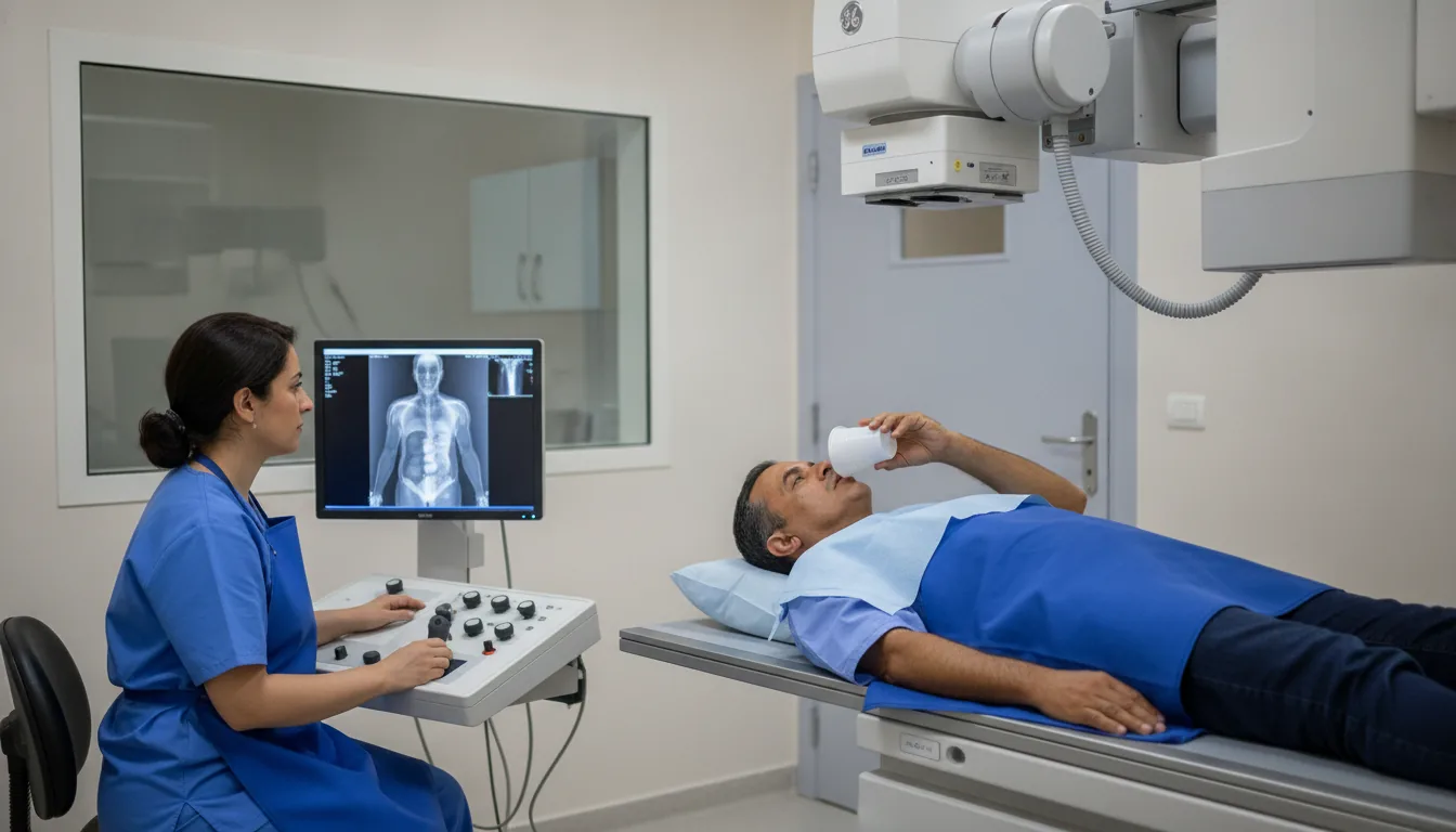
You sit next to the table with your arm flat on the plate
The elbow is straight for the front view, then bent for the side view, without forcing if it hurts
Several images may be taken from different angles, called "views": from the front, from the side (lateral), sometimes at an oblique angle
During each image you need to keep still for a few seconds; you may be asked to hold your breath
The radiographer checks that the images are sharp before you get dressed
After the examination, you can return to your usual activities straight away
Duration
About 5 to 10 minutes
Results
The images are analysed by a radiologist, who writes a report. It is given to you with the images and sent to your doctor, who will explain the results and the next steps. You will be told on the day of the examination when the results will be ready.
Precautions and contraindications
Your doctor and the radiology team will inform you in detail before the examination. Feel free to ask them your questions.
This information is general and does not replace the advice of your doctor, who chooses the examination suited to your situation.
Other examinations — Conventional radiology
Related searches
- Elbow and forearm X-rays Tunis
- Elbow and forearm X-rays Tunisia
- Elbow and forearm X-rays in Tunisia