
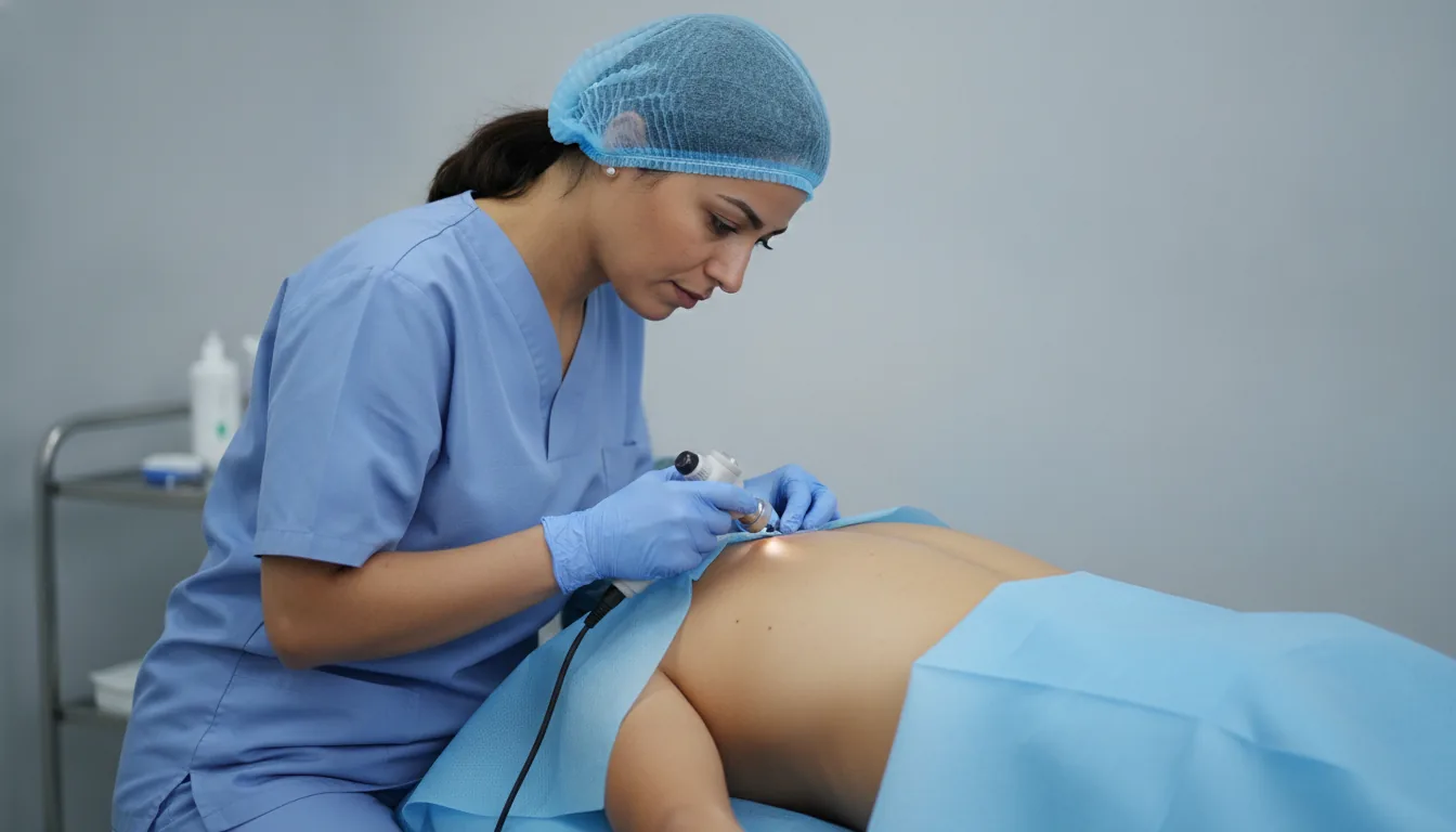
Excision of a skin lesion (mole, cyst, skin tumour)
Skin lesion excision in Tunis, Tunisia · Centre Urbain Nord
Surgical removal of a mole, a cyst or a skin tumour, with examination under the microscope.
What is it?
When is it indicated?
A mole that grows or changes colour or shape should be removed and analysed to rule out melanoma.
Skin carcinomas, common on sun-exposed areas, are most often treated by complete surgical removal.
A cyst that becomes infected or a lipoma that grows can be removed to prevent recurrence and discomfort.
A mole that catches on clothing or bleeds when shaving can be removed for comfort.
Preparation
Procedure
Marking of the area to be removed with a safety margin
Local anaesthesia of the area
Removal of the lesion
Direct closure, local flap or graft depending on the size
Lesion sent for examination under the microscope
Dressing and care instructions
Duration
20 minutes to 1 hour depending on the lesion
Aftercare & Recovery
The patient goes home straight after the procedure. Stitches are removed after one to two weeks. The result of the analysis is usually available within one to two weeks and explained at the follow-up. If a further excision or dermatological follow-up is needed, it is arranged at that point.
Risks and side effects
These risks remain rare. Your surgeon will inform you in detail before the procedure.
Coming from abroad?
The clinic supports international patients: personalised quote, stay planning, help with accommodation and transfers, communication in French, English and Arabic.
Request a Quote
Contact us to schedule a consultation or receive a personalized quote.
Request a Quote +216 36 402 000At a glance
Read also
Cosmetic Surgery in Tunisia: Planning Your Project
Cosmetic surgery is prepared well before the day of the procedure. Here are the steps of a calmly planned project in Tunis, from the first consultation to follow-up after you return home.
Read the article
Otoplasty in Tunisia: Prominent Ears, Procedure and Recovery
Otoplasty corrects prominent ears in children and adults alike. Here is how this ear surgery is prepared and performed, and what recovery looks like.
Read the articleRelated searches
- Skin lesion excision Tunis
- Skin lesion excision Tunisia
- Skin lesion excision in Tunisia