
Carotid and vertebral Doppler ultrasound (supra-aortic trunks)
Carotid Doppler in Tunis, Tunisia · Centre Urbain Nord
Carotid and vertebral Doppler ultrasound in Tunis: checks the neck arteries for plaques or narrowing, stroke work-up, without X-rays.
What is it?
When is it prescribed?
It looks for a plaque or narrowing of a carotid artery that might have caused the event.
A bruit heard with the stethoscope can mean narrowing: the test confirms and measures it.
In people at risk (smoking, diabetes, high cholesterol, high blood pressure), it assesses the neck arteries.
It follows a known narrowing over time or checks an artery after surgery.
Preparation
How the exam is performed
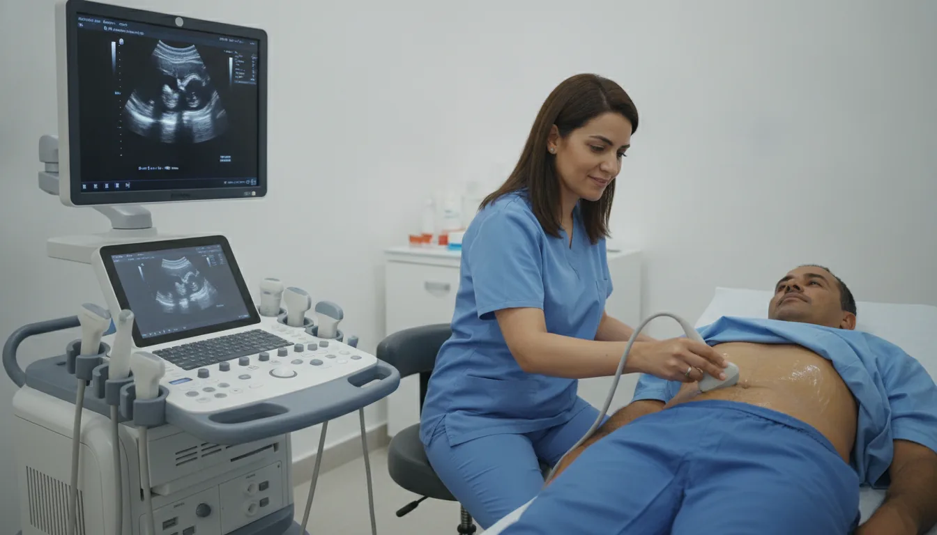
You lie on your back, with your head turned slightly away from the side being examined
A gel is applied to the skin to help the ultrasound pass through; it may feel a little cold
The probe glides along the neck, first on one side and then the other
You may hear a rhythmic whooshing sound: this is the sound of blood flowing, picked up by the Doppler
You may be asked not to talk or swallow during some measurements
The gel is wiped off at the end; you can return to your usual activities straight away
Duration
About 15 to 30 minutes
Results
The doctor performing the test (cardiologist, angiologist or radiologist, depending on the referral) analyses the images and measurements during the examination, then writes a report. The report is given to you and sent to your doctor, who will explain the results and the next steps. You will be told when to expect it on the day of the examination.
Risks and side effects
These risks remain rare. Your physician will explain them in detail before the exam.
Request a quote
Request your quote online or call us on the number below:
Request a Quote +216 36 402 034At a glance
Related searches
- Carotid Doppler Tunis
- Carotid Doppler Tunisia
- Carotid Doppler in Tunisia