
Abdominal Doppler ultrasound (portal vein, aorta, digestive vessels)
Diagnostic Testing/Cardiac/Abdominal Doppler

Abdominal Doppler ultrasound (portal vein, aorta, digestive vessels)

Abdominal Doppler in Tunis, Tunisia · Centre Urbain Nord

Abdominal Doppler ultrasound in Tunis: portal vein, aorta and digestive arteries, for cirrhosis, aneurysm or pain after meals.

What is it?

An abdominal Doppler examines the large blood vessels of the abdomen: the abdominal aorta, the portal vein (which carries blood from the bowel to the liver), the liver veins and the arteries supplying the bowel. Depending on the referral, it is performed by a cardiologist, an angiologist (a doctor specialising in blood vessels) or a radiologist. It is requested in liver disease (cirrhosis) to look for portal hypertension or a clot in the portal vein, to measure and monitor an aneurysm (widening) of the abdominal aorta, or for abdominal pain after meals, which may be caused by reduced blood supply to the bowel (mesenteric ischaemia). The test is done on an empty stomach, because gas and digestion block the ultrasound. When two non-adjacent regions need to be studied (for example the abdomen and the legs), they can be done in the same session.
Abdominal Doppler ultrasound (portal vein, aorta, digestive vessels)

When is it prescribed?

Cirrhosis or chronic liver disease

It looks for portal hypertension and checks that the portal vein is not blocked by a clot.

Abdominal aortic aneurysm

It measures the diameter of the aorta and allows a widening to be monitored over time.

Abdominal pain after meals, weight loss

It looks for narrowing of the bowel arteries (mesenteric ischaemia).

Before or after a procedure on the liver or blood vessels

It checks the blood flow in the vessels concerned.

Preparation

1Do not eat for about 6 hours beforehand (no food, no smoking, no chewing gum; a little still water is allowed), unless told otherwise: gas in the bowel blocks the ultrasound
2Take your usual medicines with a little water, unless told otherwise
3Wear clothes that are easy to remove or roll up over the area to be examined
4Bring your blood test results (liver tests) and previous ultrasound scans
5Bring your referral, previous tests (Doppler ultrasound, CT scan, MRI, angiography) and any relevant reports

How the exam is performed

1

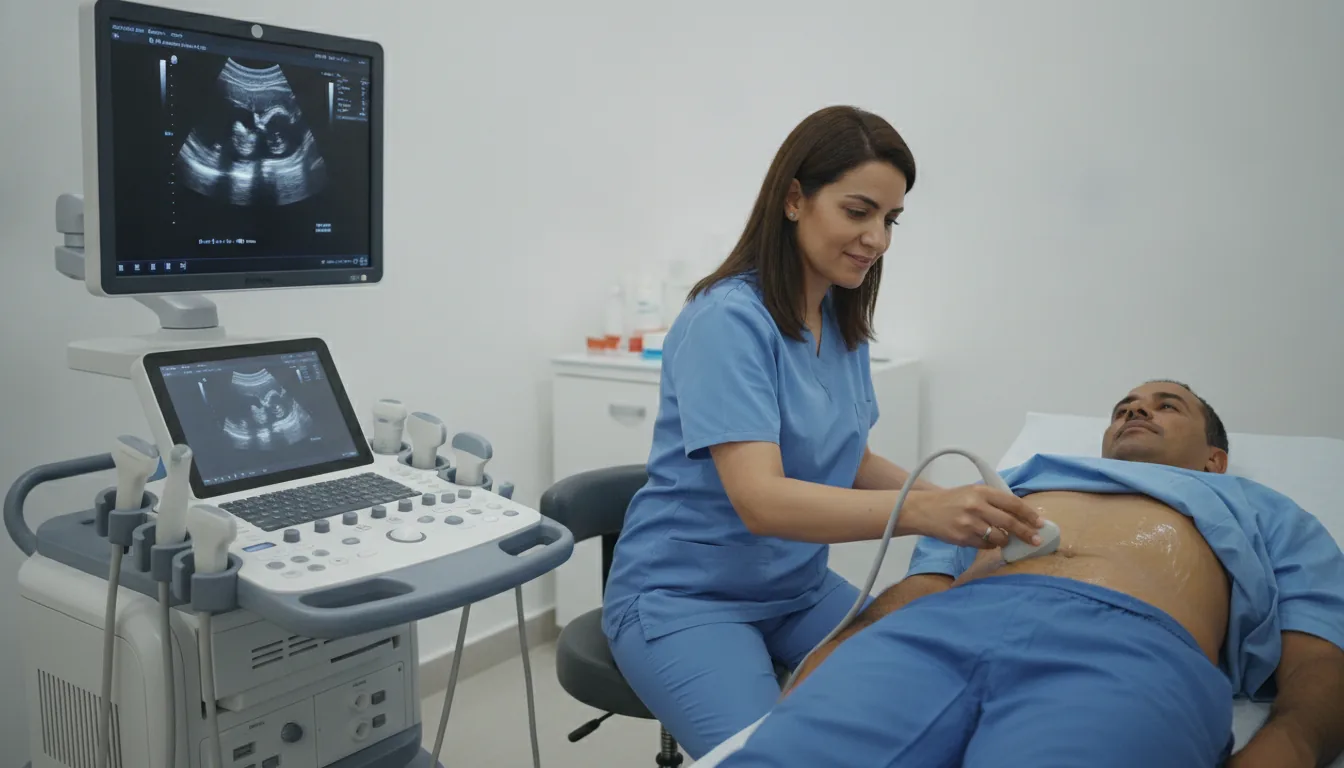
You lie on your back, and possibly on your side

2

A gel is applied to the skin to help the ultrasound pass through; it may feel a little cold

3

The probe is moved over the abdomen, pressing at times to push the gas aside

4

You may be asked to breathe in deeply and hold your breath for a few seconds

5

You may hear a rhythmic whooshing sound: this is the sound of blood flowing, picked up by the Doppler

6

The gel is wiped off at the end; you can return to your usual activities straight away

Duration

About 20 to 40 minutes

Results

The doctor performing the test (cardiologist, angiologist or radiologist, depending on the referral) analyses the images and measurements during the examination, then writes a report. The report is given to you and sent to your doctor, who will explain the results and the next steps. You will be told when to expect it on the day of the examination.

Risks and side effects

No X-rays: Doppler ultrasound uses only ultrasound waves
No injection or contrast agent
Painless; the pressure of the probe on the abdomen may be a little uncomfortable
Can be done during pregnancy, as ultrasound is not radiation

These risks remain rare. Your physician will explain them in detail before the exam.

Request a quote

Request your quote online or call us on the number below:

Request a Quote +216 36 402 034

At a glance

Duration: About 20 to 40 minutes
Preparation: Required

Related searches

  • Abdominal Doppler Tunis
  • Abdominal Doppler Tunisia
  • Abdominal Doppler in Tunisia
Request a quoteBook an appointment